Home
Images
Vectors
Videos
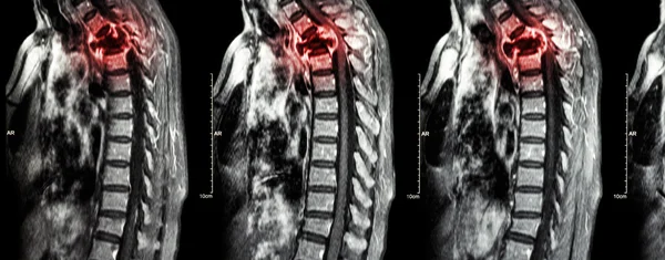
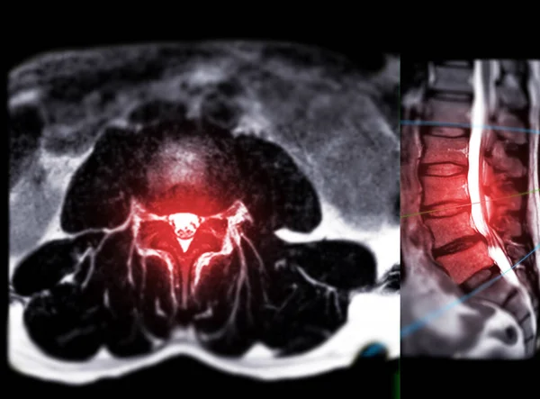
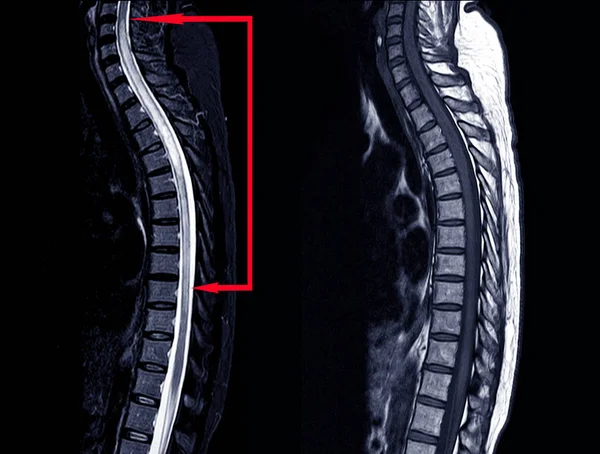
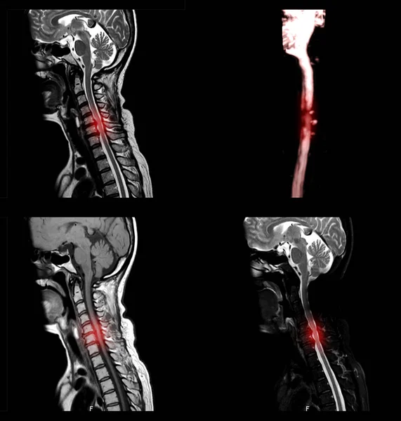
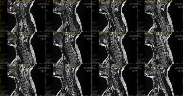
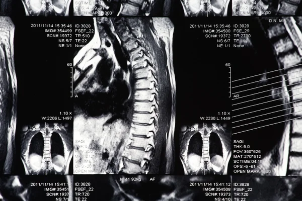
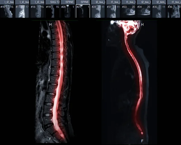
Stock image MRI of whole spine T2W sagittal plane for diagnostic Spinal Cord Compression.
Published:
Jul.07, 2021 06:04:23
Author:
samunella
Views:
6
Downloads:
0
File type:
image
/
jpg
File size:
0.99 MB
Orginal size:
2560
x
2048
px
Available sizes:
S
(500 x 400)
M
(1000 x 800)
L
(2000 x 1600)
XL
(2560 x 2048)
EL0
(2560 x 2048)
Level:
beginner
Download Image
Image topics:
Image
Facet
Health
Medicine
Illness
Medical
Cord
Hospital
Patient
Neck
Disease
Pain
Compression
Column
Disc
Muscle
Spine
Magnetic
Skeleton
Fracture
Diagnosis
Examination
Spinal
Vertebra
Brain
Imaging
Surgery
Accident
Injury
Scan
Resonance
Radiology
Orthopedic
Nerve
Thoracic
Ct
Degenerative
Cervical
Lumbar
Vertebrae
Spinal Cord
Spinal Column
Cervical Spine
Paresthesia
Spinal Injury
Sagittal Plane
Myelogram
Mri Myelogram
Whole Spine
Mri Whole Spine
Similar stock images
MRI Of Thoracic Spine Moderate Pathologic Compression Fracture Of T9 Vertebral Body With Paravertebral Abscess At T9-10.
7000 × 4205
Spine Metastasis ( Cancer Spread To Thoracic Spine ) ( MRI Of Cervical And Thoracic Spine : Show Thoracic Spine Metastasis And Compress Spinal Cord ( Myelopathy ) ) ( Sagittal Plane )
5127 × 2014
MRI L-S Spine Or Lumbar Spine Axial T2W View With Sagittal Plane For Diagnosis Spinal Cord Compression.
3032 × 2240
MRI Scan Of Lower Back Region.
2627 × 3500
MRI OF THORACIC SPINE HISTORY: A 53-year-old Woman, Presented Wi
5144 × 3900
A Sagittal View Of MRI C-spine Or Magnetic Resonance Image Of Cervical Spine Showing Spondylosis Causing Cervical Spondylotic Myelopathy And Compression Fracture.
2835 × 2976
Set Of Sagittal MRI Scans Of Neck Area Of Caucasian 34 Years Old Male With Bilateral Paramedial Extrusion Of The C6-C7 Segment With Radiculopathy
3840 × 2019
X-ray Photograph
3000 × 2000