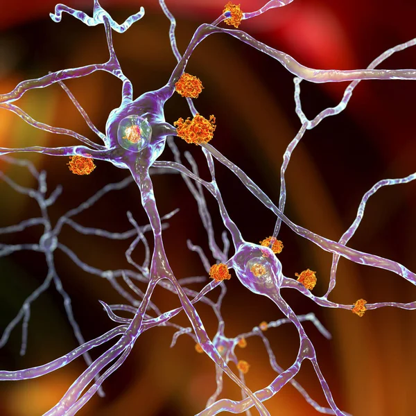
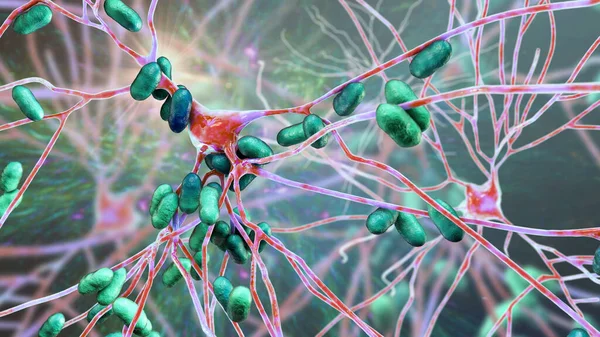
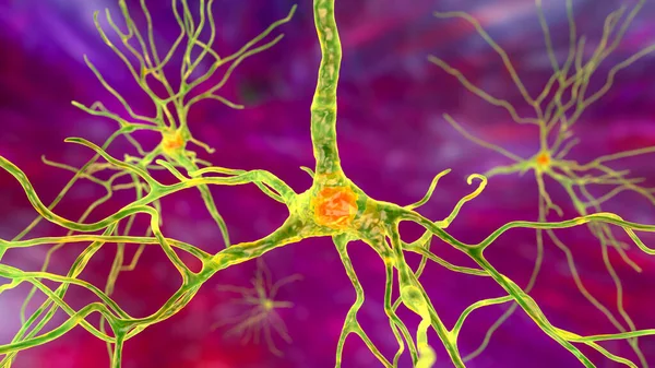
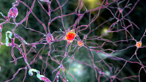
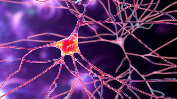
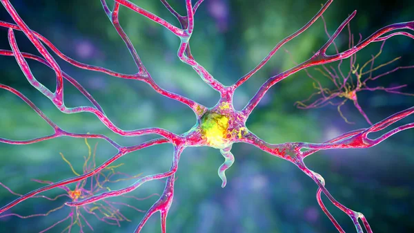
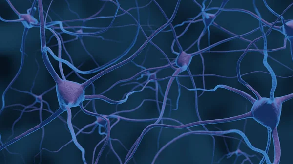
Stock image Neurons, 3D illustration showing brain cells located in the temporal cortex of the human brain in Brodmann area 20. They are involved in high-level visual processing and recognition memory

Published: Dec.03, 2020 14:34:00
Author: katerynakon
Views: 71
Downloads: 2
File type: image / jpg
File size: 8.38 MB
Orginal size: 7200 x 4050 px
Available sizes:
Level: silver