
Stock image Brodmann
Human Brain With Highlighted Fusiform Gyrus, Or Medial Occipitotemporal Gyrus, 3D Illustration. It Is Associated With Various Neural Pathways Related To Recognition, And Also To Synesthesia, Dyslexia, And Prosopagnosia
Image, 4.67MB, 6000 × 4000 jpg
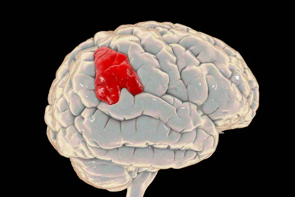
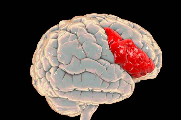
Human Brain With Highlighted Supramarginal Gyrus, 3D Illustration. It Is Involved In Perception Of Language, Space And Limbs Location, Identifying Postures And Gestures Of Other People
Image, 5.74MB, 6000 × 4000 jpg
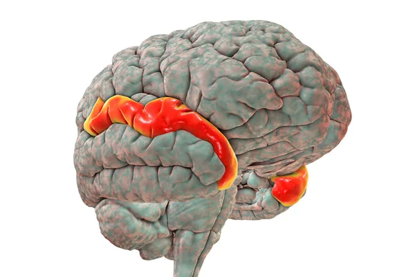
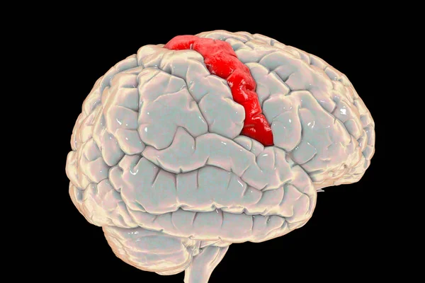
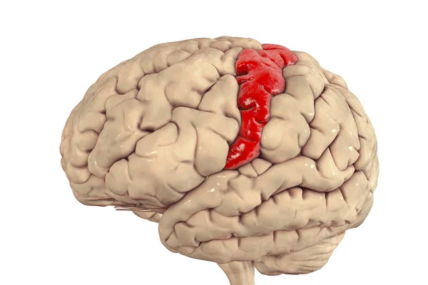
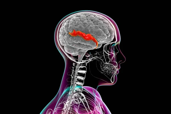
Human Brain With Highlighted Superior Temporal Gyrus, 3D Illustration. It Is Located In The Temporal Lobe, Contains The Auditory Cortex, Is Responsible For The Sensation Of Sound And The Processing Of Speech
Image, 5.62MB, 6000 × 4000 jpg
Human Brain With Highlighted Precentral Gyrus, Side View, 3D Illustration. It Is Located In The Posterior Frontal Lobe And Is The Site Of The Primary Motor Cortex, The Brodmann Area 4.
Image, 5.54MB, 6000 × 4000 jpg
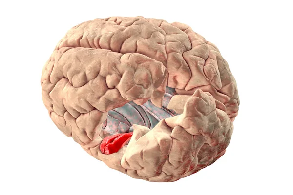
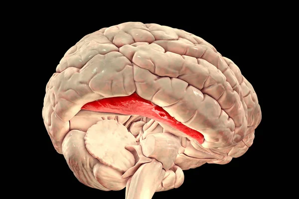
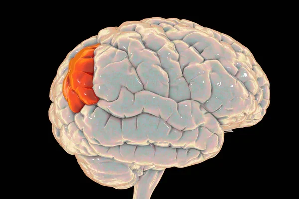
Human Brain With Highlighted In Red Transverse Temporal Gyri, Which Are Part Of Primary Auditory Cortex, 3D Illustration. Left Precentral And Postcentral Gyri Are Deleted For Better Visualization
Image, 6.26MB, 6230 × 4153 jpg

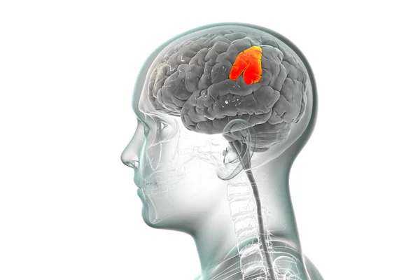
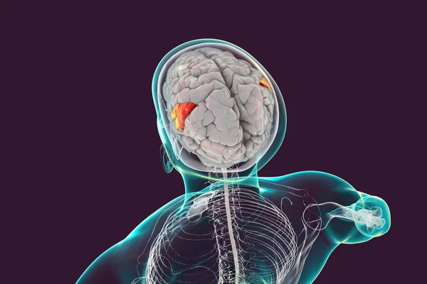
Human Brain In Body With Highlighted Supramarginal Gyrus, 3D Illustration. It Is Involved In Perception Of Language, Space And Limbs Location, Identifying Postures And Gestures Of Other People
Image, 5.29MB, 6000 × 4000 jpg
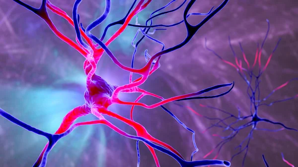
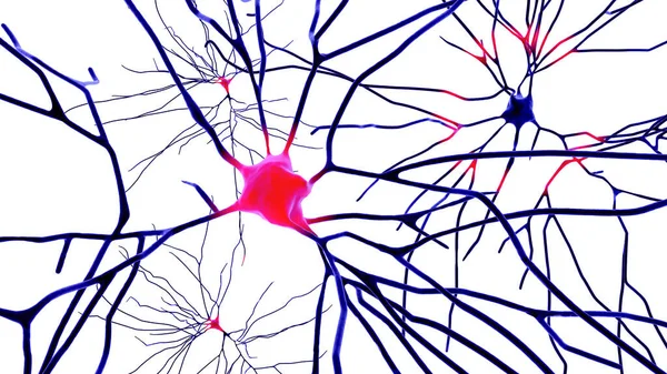
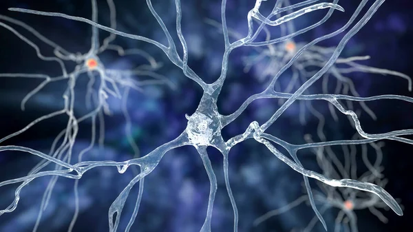
Neurons, 3D Illustration Showing Brain Cells Located In The Precentral Gyrus Of The Frontal Cortex Of The Human Brain. They Control Movements Of The Contralateral Side Of The Body
Image, 7.01MB, 7200 × 4050 jpg
Human Brain With Highlighted Precentral Gyrus, 3D Illustration. It Is Located In The Posterior Frontal Lobe And Is The Site Of The Primary Motor Cortex, The Brodmann Area 4.
Image, 4.54MB, 6100 × 4066 jpg
Human Brain In The Body With Highlighted Precentral Gyrus, 3D Illustration. It Is Located In The Posterior Frontal Lobe And Is The Site Of The Primary Motor Cortex, The Brodmann Area 4.
Image, 3.56MB, 6000 × 4000 jpg
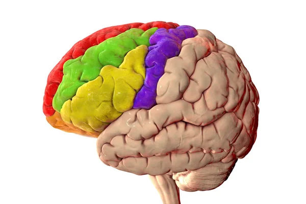
Human Brain With Highlighted Frontal Gyri, Superior (red), Middle (green), Inferior (yellow), Precentral (blue), 3D Illustration
Image, 5.09MB, 6000 × 4000 jpg
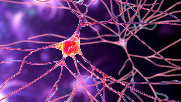
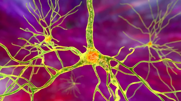
Neurons, 3D Illustration Showing Brain Cells Located In The Temporal Cortex Of The Human Brain In Brodmann Area 20. They Are Involved In High-level Visual Processing And Recognition Memory
Image, 11.76MB, 7200 × 4050 jpg
Human Brain With Highlighted Angular Gyrus, 3D Illustration. It Is Located In The Parietal Lobe, Is Involved Into Language, Number Processing, Spatial Cognition, Attention, And Theory Of Mind
Image, 5.57MB, 6000 × 4000 jpg
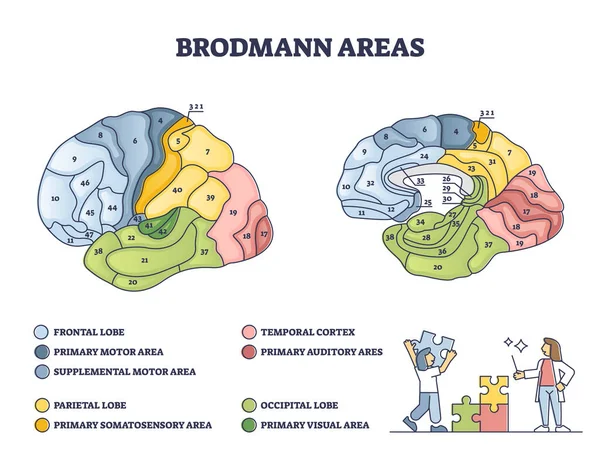
Brodmann Areas Map As Brain Region Zones Of Cerebral Cortex Outline Diagram
Vector, 6.18MB, 4800 × 3733 eps
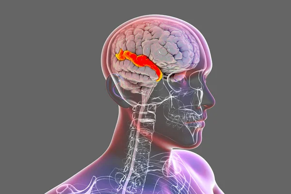
Human Brain In Body With Highlighted Superior Temporal Gyrus, 3D Illustration. It Is Located In The Temporal Lobe, Contains The Auditory Cortex, And Is Responsible For The Processing Of Speech
Image, 5.33MB, 6000 × 4000 jpg
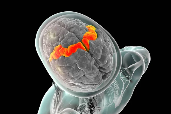
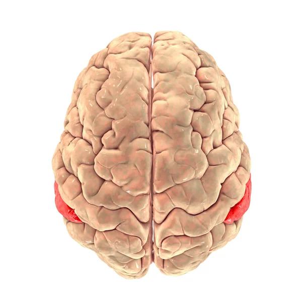
Human Brain With Highlighted Angular Gyrus, Top View, 3D Illustration. It Is Located In The Parietal Lobe, Is Involved Into Language, Number Processing, Spatial Cognition, Attention, Theory Of Mind
Image, 4.59MB, 5000 × 5000 jpg
Human Brain With Highlighted Inferior Frontal Gyrus, 3D Illustration. It Is A Part Of The Prefrontal Cortex And The Location Of Broca's Area, Involved In Language Processing And Speech Production
Image, 6.07MB, 6000 × 4000 jpg
Neurons Isolated On White Background, 3D Illustration Showing Brain Cells Located In The Temporal Cortex Of The Human Brain In Brodmann Area 20. They Are Involved In High-level Visual Processing
Image, 5.9MB, 7200 × 4050 jpg

Neurons, 3D Illustration Showing Brain Cells Located In The Temporal Cortex Of The Human Brain In Brodmann Area 20. They Are Involved In High-level Visual Processing And Recognition Memory
Image, 8.38MB, 7200 × 4050 jpg
Neurons, 3D Illustration Showing Brain Cells Located In The Precentral Gyrus Of The Frontal Cortex Of The Human Brain. They Control Movements Of The Contralateral Side Of The Body
Image, 8.34MB, 7200 × 4050 jpg

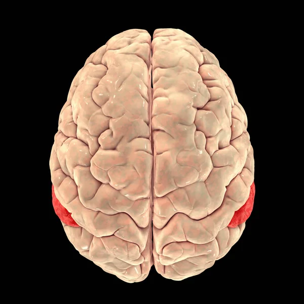
Human Brain With Highlighted Superior Parietal Lobule, 3D Illustration
Image, 4.74MB, 6208 × 4138 jpg

Human Brain With Highlighted Temporal Gyri, 3D Illustration. Superior Temporal Gyrus (orange), Middle (blue), And Inferior (red). They Are Involved In Processing Auditory Information And Encoding Of Memory
Image, 5.74MB, 6000 × 4000 jpg

Neurons, 3D Illustration Showing Brain Cells Located In The Temporal Cortex Of The Human Brain In Brodmann Area 20. They Are Involved In High-level Visual Processing And Recognition Memory
Image, 7.24MB, 7200 × 4050 jpg
Human Brain In Body With Highlighted Supramarginal Gyrus, 3D Illustration. It Is Involved In Perception Of Language, Space And Limbs Location, Identifying Postures And Gestures Of Other People
Image, 2.57MB, 6000 × 4000 jpg

Neurons, 3D Illustration Showing Brain Cells Located In The Precentral Gyrus Of The Frontal Cortex Of The Human Brain. They Control Movements Of The Contralateral Side Of The Body
Image, 4MB, 7200 × 4050 jpg

Neurons, 3D Illustration Showing Brain Cells Located In The Temporal Cortex Of The Human Brain In Brodmann Area 20. They Are Involved In High-level Visual Processing And Recognition Memory
Image, 8.08MB, 7200 × 4050 jpg

Human Brain With Highlighted Temporal Lobe And Close-up View Of Pyramidal Neurons Found In Temporal Cortex, 3D Illustration
Image, 8.78MB, 7802 × 4388 jpg
Human Brain In Body With Highlighted Superior Temporal Gyrus, 3D Illustration. It Is Located In The Temporal Lobe, Contains The Auditory Cortex, And Is Responsible For The Processing Of Speech
Image, 3.6MB, 6000 × 4000 jpg
Human Brain With Highlighted Supramarginal Gyrus, Top View, 3D Illustration. It Is Involved In Perception Of Language, Space And Limbs Location, Identifying Postures And Gestures Of Other People
Image, 4.95MB, 5000 × 5000 jpg

Human Brain With Highlighted Temporal Lobe And Close-up View Of Pyramidal Neurons Found In Temporal Cortex, 3D Illustration
Image, 8MB, 6000 × 4000 jpg
Neurons, 3D Illustration Showing Brain Cells Located In The Precentral Gyrus Of The Frontal Cortex Of The Human Brain. They Control Movements Of The Contralateral Side Of The Body
Image, 8.69MB, 7200 × 4050 jpg
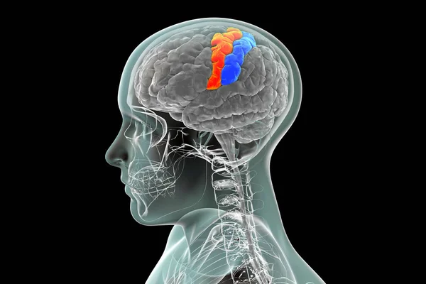
Human Brain With Highlighted Precentral And Postcentral Gyri, 3D Illustration. The Sites Of Primary Motor (precentral Gyrus) And Somatosensory (postcentral Gyrus) Cortex
Image, 3.12MB, 6000 × 4000 jpg
Human Brain In Body With Highlighted Supramarginal Gyrus, 3D Illustration. It Is Involved In Perception Of Language, Space And Limbs Location, Identifying Postures And Gestures Of Other People
Image, 5.35MB, 6000 × 4000 jpg
Page 1 >> Next