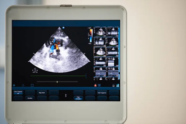
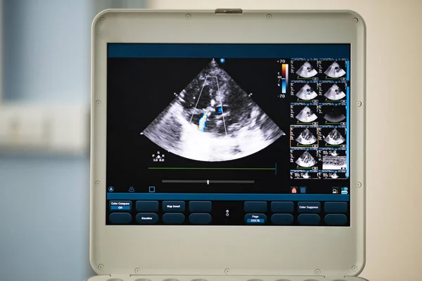
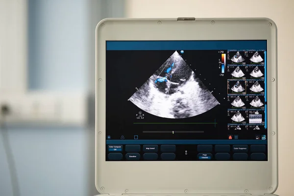
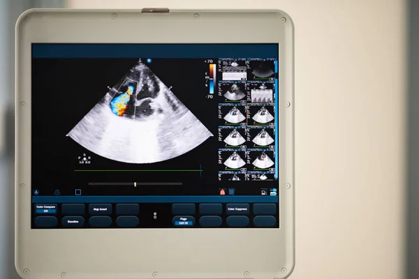
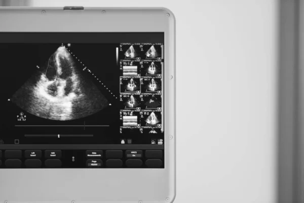
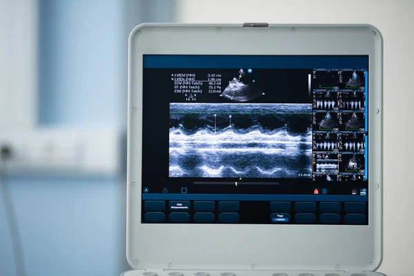
Stock image On the screen of the ultrasound apparatus, the scan of the left ventricle of the heart in the position for measuring the ejection fraction by Teyolz.

Published: Jul.23, 2018 09:02:51
Author: Faustasyan
Views: 11
Downloads: 0
File type: image / jpg
File size: 14.44 MB
Orginal size: 6000 x 4005 px
Available sizes:
Level: beginner