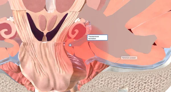
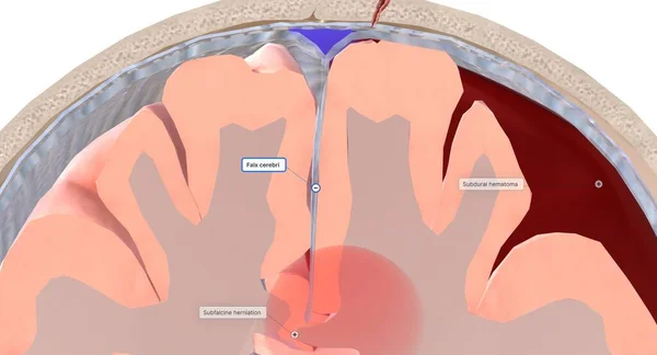
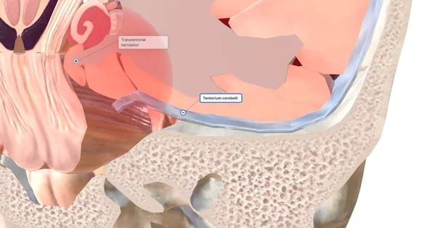
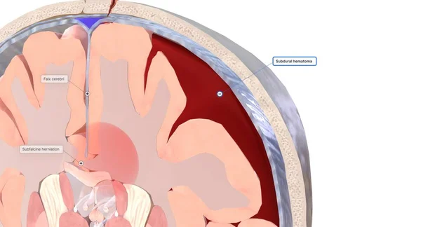
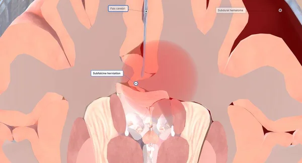
Stock image One common type of brain herniation is characterized by brain tissue moving underneath the middle fold of dura (falx cerebri). This type is called a subfalcine brain herniation. 3D rendering
Published: Jun.19, 2023 10:20:27
Author: scienceanm
Views: 0
Downloads: 0
File type: image / jpg
File size: 10.02 MB
Orginal size: 7258 x 3920 px
Available sizes:
Level: bronze