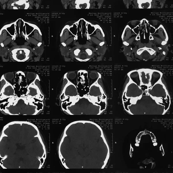
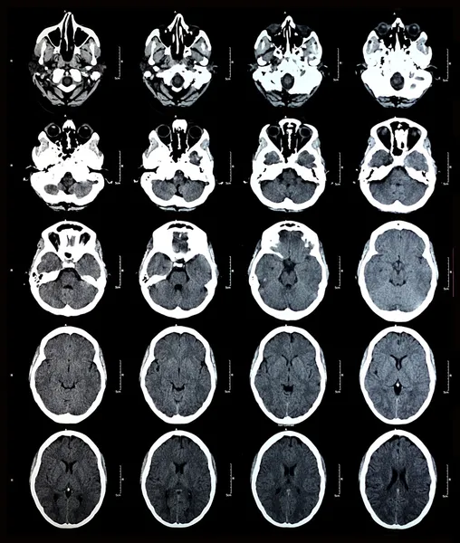
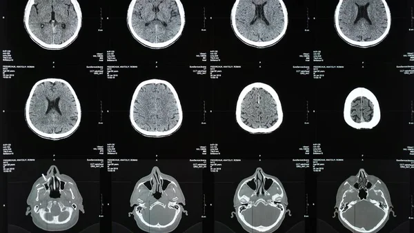
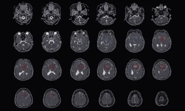
Stock image Perfusion CT scan image of the brain 3d rendering image analysing on the screen monitor.

Published: Dec.23, 2019 11:44:46
Author: samunella
Views: 18
Downloads: 0
File type: image / jpg
File size: 2.56 MB
Orginal size: 3284 x 2200 px
Available sizes:
Level: beginner