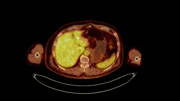
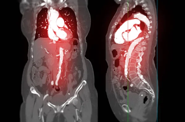
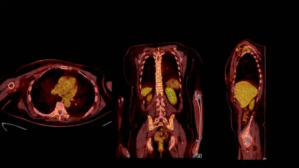
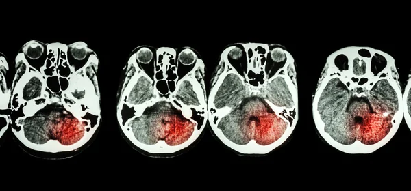
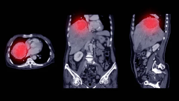
Stock image PET CT Scan fusion image It provides detailed images by merging metabolic activity from PET with anatomical information from CT scans.
Published: Dec.28, 2023 12:34:43
Author: samunella
Views: 1
Downloads: 0
File type: image / jpg
File size: 3.21 MB
Orginal size: 3840 x 2160 px
Available sizes:
Level: beginner
Similar stock images
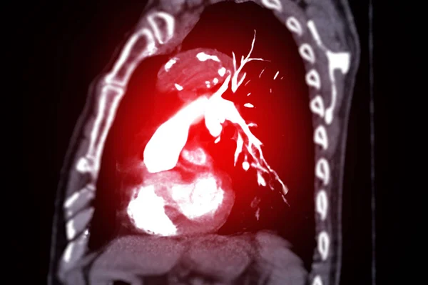
CTPA Or CTA Pulmonary Artery For Diagnostic Pulmonary Embolism (PE) , Lung Cancer And Covid-19. .
4866 × 3240