Home
Images
Vectors
Videos
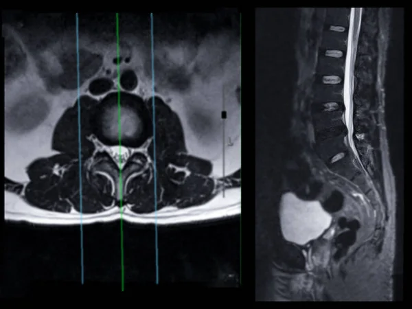
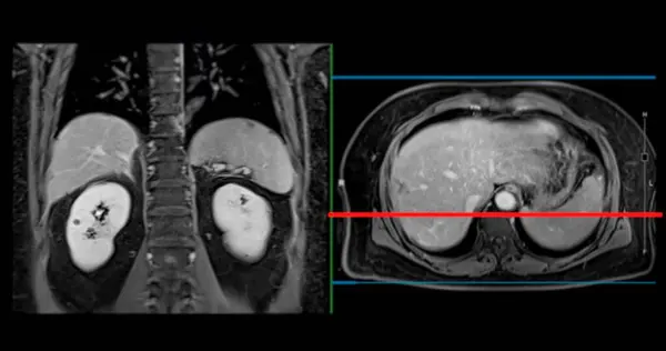
Stock image PET MRI of the liver in liver cancer provides precise imaging, aiding in tumor detection, staging, and treatment planning.
Published:
May.02, 2024 08:48:14
Author:
samunella
Views:
0
Downloads:
0
File type:
image
/
jpg
File size:
0.49 MB
Orginal size:
5096
x
2687
px
Available sizes:
S
(500 x 264)
M
(1000 x 527)
L
(2000 x 1055)
XL
(5096 x 2687)
EL0
(5096 x 2687)
Level:
beginner
Download Image
Image topics:
Health
Illness
Medical
Body
Abdomen
Disease
Cholesterol
Post
Diagnosis
Imaging
Digestive
Scan
Obstruction
Abdominal
Liver
Duct
Phase
Tumor
Inflammation
Hypoglycemia
Bladder
Pancreas
Venous
Polyps
Duodenum
Hepatic
Malignancy
Abscess
Carcinoma
MRI Scan
Abdominal Pain
Magnetic Resonance Imaging
Adenocarcinoma
Cholecystitis
Liver Disorders
Carcinomatosis
Hepatomegaly
Mri Abdomen
Mri Upper Abdomen
Inflection Of The Gallbladder
Gallbladder Deformity
Gallbladder Polyps
Gallbladder Diseases
Abdominal Diagnosis
Upper Abdominal Region
Organ Health Assessment
Benign Lesions
Mri Of Upper Abdomen
Similar stock images
PET MRI Of The Liver In Liver Cancer Provides Precise Imaging, Aiding In Tumor Detection, Staging, And Treatment Planning.
5096 × 2687
PET MRI Of The Liver In Liver Cancer Provides Precise Imaging, Aiding In Tumor Detection, Staging, And Treatment Planning.
5096 × 2687
MRI Of The Upper Abdomen Is A Non-invasive Imaging Technique Providing Detailed Visuals Of Organs Like The Liver, Pancreas, And Kidneys In Case Normal Study.
5096 × 2687
MRI Of The Upper Abdomen Is A Non-invasive Imaging Technique Providing Detailed Visuals Of Organs Like The Liver, Pancreas, And Kidneys In Case Normal Study.
5096 × 2687
MRI Of The Upper Abdomen Is A Non-invasive Imaging Technique Providing Detailed Visuals Of Organs Like The Liver, Pancreas, And Kidneys In Case Normal Study.
5096 × 2687
MRI Of The Upper Abdomen Is A Non-invasive Imaging Technique Providing Detailed Visuals Of Organs Like The Liver, Pancreas, And Kidneys In Case Normal Study.
5601 × 3258
MRI L-S Spine Or Lumbar Spine Axial T2W View With Sagittal Plane For Diagnosis Spinal Cord Compression.
4000 × 3000
Real-time 3D Constructions From Patient Images, Scanner Processed For 3D Printing.
5616 × 3744