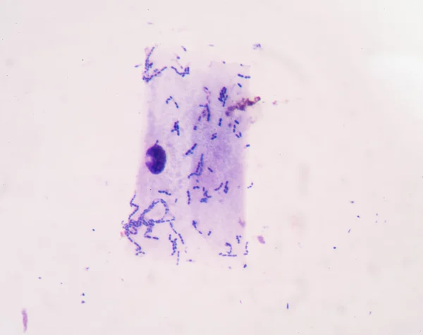
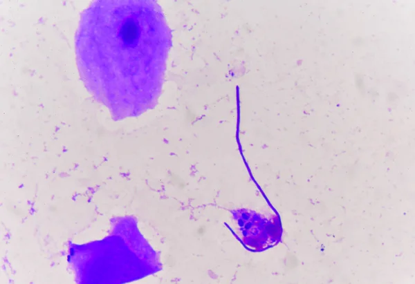
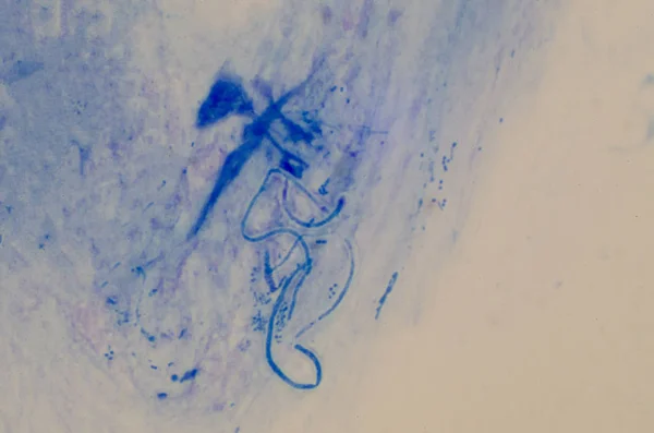
Stock image Pseudohyphae in gram stain method.
Published: Jul.24, 2019 10:44:48
Author: toeytoey
Views: 116
Downloads: 2
File type: image / jpg
File size: 10.2 MB
Orginal size: 4928 x 3264 px
Available sizes:
Level: bronze