
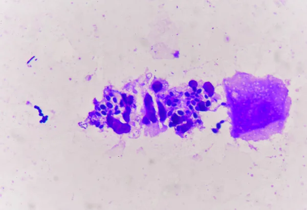
Stock image Hyphal
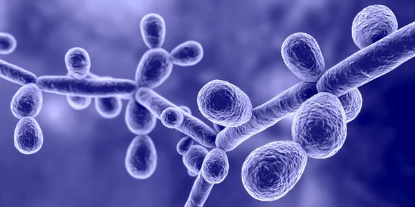
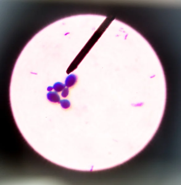
Candida Tropicalis Yeasts, Microscopic Fungi That Cause Infections In Immunocompromised Patients. Scientific 3D Illustration Showing Pseudohyphae And Blastoconidia Formed Singly Or In Small Groups
Image, 7.09MB, 8000 × 4000 jpg

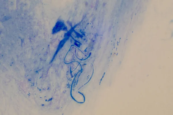
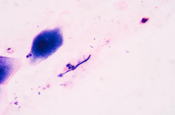
Histoplasma Capsulatum, A Parasitic, Yeast-like Dimorphic Fungus That Can Cause Lung Infection Histoplasmosis. A 3D Illustration Depicts A Mycelial Form Found In Soil Enriched By Animal Excrement.
Image, 6.99MB, 7200 × 4050 jpg


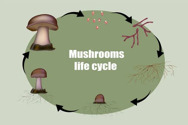
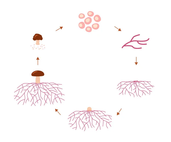
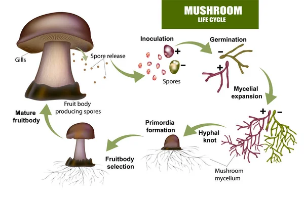
Mushroom Life Cycle Stages, Growth Mycelium From Spore. Spore Germination, Mycelial Expansion And Formation Hyphal Knot. Vector
Vector, 0.76MB, 9000 × 3000 eps
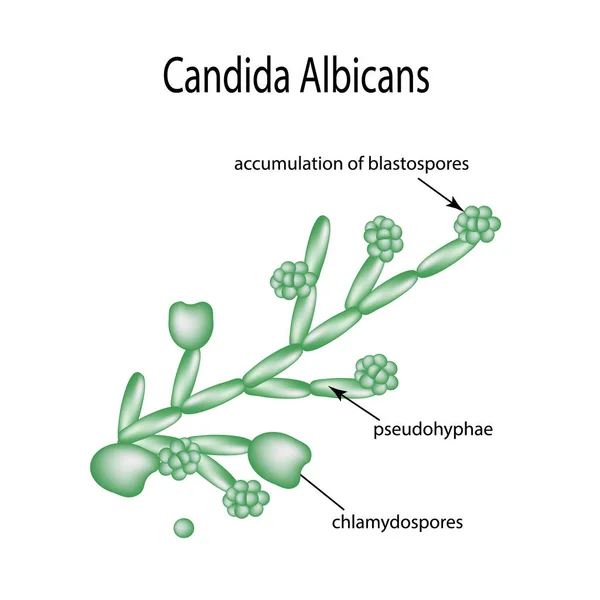
Structure Of Candida Albicans. Infographics. Vector Illustration On Isolated Background
Vector, 2.17MB, 5000 × 5000 eps
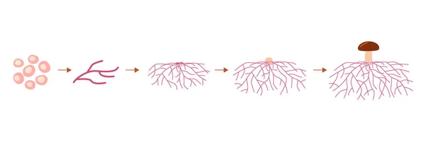
Mushroom Life Cycle Stages, Growth Mycelium From Spore. Spore Germination, Mycelial Expansion And Formation Hyphal Knot. Vector Illustration
Vector, 0.46MB, 6000 × 5000 eps
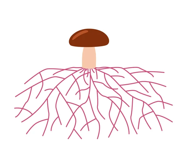
Mushroom Life, Growth Mycelium From Spore. Spore Germination, Mycelial Expansion And Formation Hyphal Knot. Vector Illustration
Vector, 0.76MB, 6000 × 5000 eps


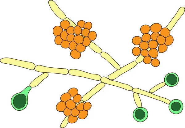
Candida Albicans Yeasts, The Causative Agent Of Candidiasis. Scientific Vector Illustration Showing Pseudohyphae (yellow), Blastoconidia (orange) And Chlamydospores (green)
Vector, 5.21MB, 5665 × 3910 eps


3d Rendering Of Candida Albicans, It Is An Opportunistic Pathogenic Yeast That Is A Common Member Of The Human Gut Flora
Image, 0.87MB, 3840 × 2160 jpg

3d Rendering Of Candida Albicans, It Is An Opportunistic Pathogenic Yeast That Is A Common Member Of The Human Gut Flora
Image, 0.93MB, 3840 × 2160 jpg

3d Rendering Of Candida Albicans, It Is An Opportunistic Pathogenic Yeast That Is A Common Member Of The Human Gut Flora
Image, 0.8MB, 3840 × 2160 jpg
Page 1 >> Next