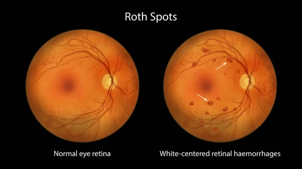
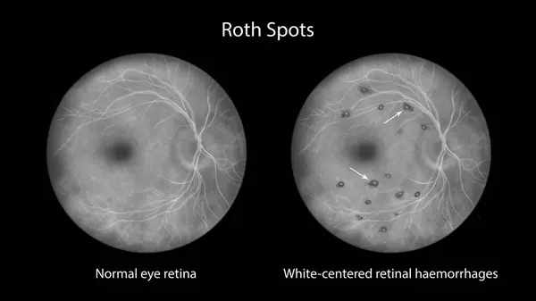
Stock image Roth spots in the retina seen during ophthalmoscopy, a 3D illustration showing white-centered retinal hemorrhages with surrounding hemorrhagic rings.

Published: Sep.26, 2023 14:24:42
Author: katerynakon
Views: 1
Downloads: 1
File type: image / jpg
File size: 19.32 MB
Orginal size: 11738 x 6603 px
Available sizes:
Level: silver