
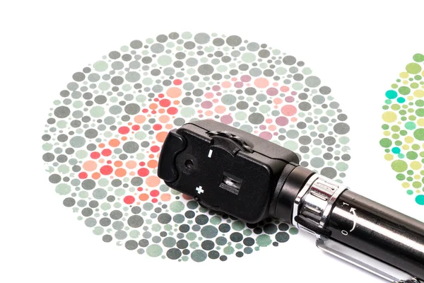
Stock image Ophthalmoscopy


Proliferative Diabetic Retinopathy, Illustration Showing Neovascularization In The Disk And Macula Edema. Abnormal Finding On Fundoscopic Examination Of The Eye Retina In Diabetes Mellitus
Image, 2.85MB, 5000 × 5000 jpg


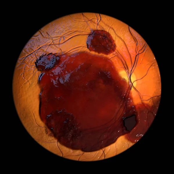
Medical 3D Illustration Of A Subretinal Hemorrhage Observed During Ophthalmoscopy, Revealing A Dark, Irregular Hemorrhage Beneath The Retinal Layers.
Image, 9.37MB, 5352 × 5352 jpg
Non-proliferative Diabetic Retinopathy, Illustration Showing Cotton Wool Spots As Fluffy Dark Patches, Abnormal Finding On Funduscopic Examination Of The Eye Retina In Diabetes Mellitus, Fluorescein Angiography
Image, 2.72MB, 5000 × 5000 jpg

A Prepapillary Vascular Loop On The Retina, As Observed During Ophthalmoscopy In Fluorescein Angiogram, An Illustration Showcasing The Looping Blood Vessels Around The Optic Disc.
Image, 2.77MB, 5000 × 5000 jpg
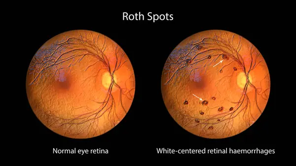
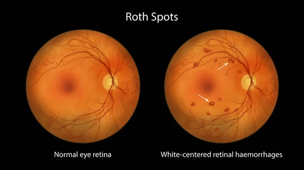
Roth Spots In The Retina Seen During Ophthalmoscopy, A 3D Illustration Showing White-centered Retinal Hemorrhages With Surrounding Hemorrhagic Rings.
Image, 19.32MB, 11738 × 6603 jpg

Non-proliferative Diabetic Retinopathy, 3D Illustration Showing Normal Eye Retina And Retina With Hard Exudates (irregularly Shaped Yellow Spots)
Image, 25.27MB, 11738 × 6603 jpg
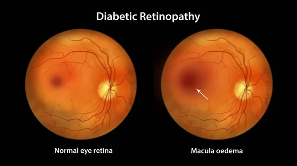
Diabetic Macular Edema (DME), Illustration Showing Normal Eye Retina And Retina With Macula Edema. Fundoscopic Examination Of The Eye Retina In Diabetes Mellitus
Image, 6.24MB, 11738 × 6603 jpg
Proliferative Diabetic Retinopathy, Illustration Showing Neovascularization In The Disk And Other Sites, And Macula Edema. Fundoscopic Examination Of The Eye Retina In Diabetes Mellitus
Image, 2.9MB, 5000 × 5000 jpg

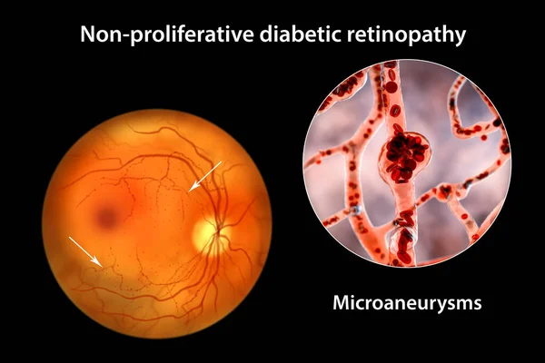
Non-proliferative Diabetic Retinopathy, 3D Illustration Showing Multiple Microaneurysms On The Eye Retina And Closeup View Of Microaneurysms, Microscopic Buldges In The Artery Walls Filled With Blood
Image, 11.11MB, 10431 × 6954 jpg


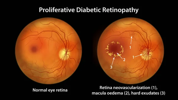
Proliferative Diabetic Retinopathy, Illustration Showing Neovascularization In The Disk And Other Sites, Macula Edema And Hard Exudates. Fundoscopic Examination Of The Eye Retina In Diabetes Mellitus
Image, 3.05MB, 5000 × 5000 jpg
Proliferative Diabetic Retinopathy, Illustration Showing Neovascularization In The Disk And Other Sites, Macula Edema And Hard Exudates. Fundoscopic Examination Of The Eye Retina In Diabetes Mellitus
Image, 6.95MB, 11738 × 6603 jpg

Non-proliferative Diabetic Retinopathy, Illustration Showing Normal Eye Retina And Retina With Hard Exudates, Microaneurysms, Dot Haemorrhages, Flame-shaped And Splinter Retinal Haemorrhages
Image, 6.89MB, 11738 × 6603 jpg
Non-proliferative Diabetic Retinopathy, Illustration Showing IRMAs (intraretinal Microvascular Abnormalities) As Small Vessels With Abnormal Branching Or Dilatation In Ischaemic Areas
Image, 2.68MB, 5000 × 5000 jpg
Non-proliferative Diabetic Retinopathy, 3D Illustration Showing Multiple Microaneurysms On The Eye Retina And Closeup View Of Microaneurysms, Microscopic Buldges In The Artery Walls Filled With Blood
Image, 10.46MB, 10431 × 6954 jpg
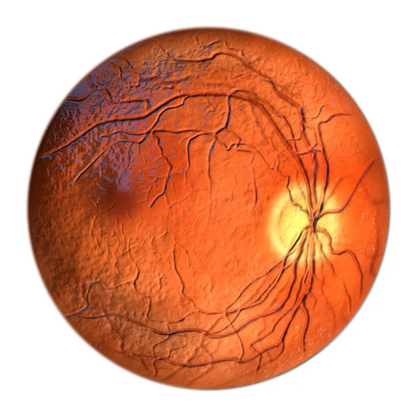
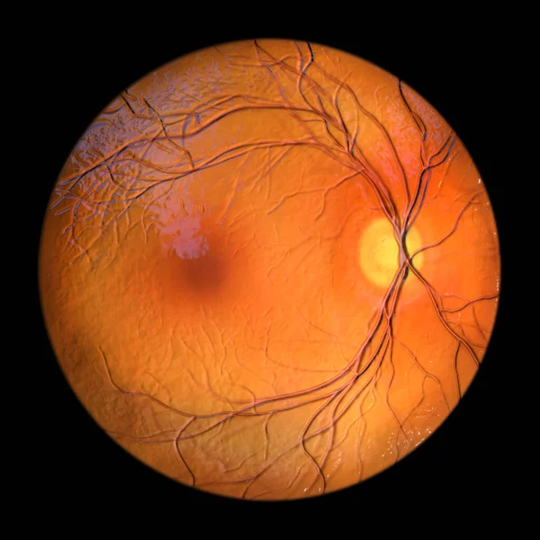
Normal Eye Retina, Ophthalmoscope View, Scientific 3D Illustration Showing Optic Disk, Blood Vessels, Macula And Fovea
Image, 13.02MB, 5352 × 5352 jpg

Direct Ophthalmoscopy. Retinal Examination. Fundoscopy. Child Vision Test In Clinic
Image, 7.53MB, 4403 × 3406 jpg
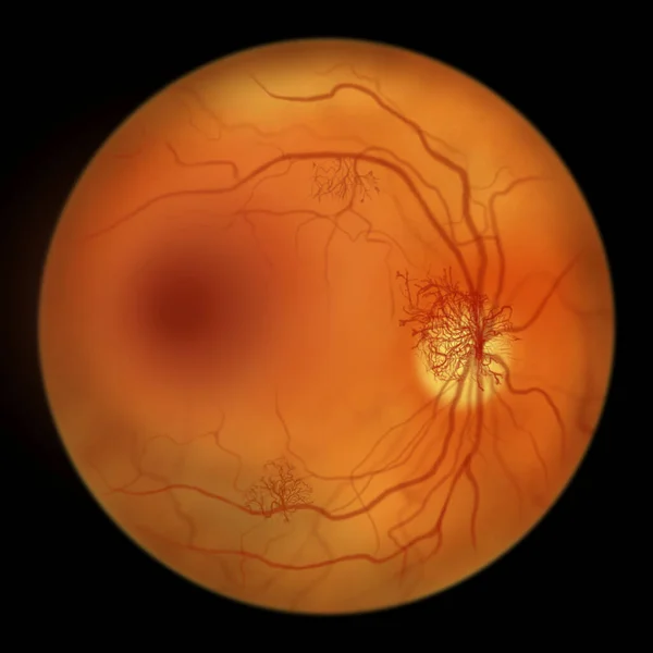
Proliferative Diabetic Retinopathy, Illustration Showing Neovascularization (formation Of New Vessels) In The Optic Disk. Fundoscopic Examination Of The Eye Retina In Diabetes Mellitus
Image, 2.81MB, 5000 × 5000 jpg

Diabetic Macular Edema (DME). Diabetic Retinopathy, Illustration Showing Macula Edema, Abnormal Finding On Fundoscopic Examination Of The Eye Retina In Diabetes Mellitus
Image, 2.72MB, 5000 × 5000 jpg


Diabetic Retinopathy, 3D Illustration Showing Macula Edema, Optic Disk Edema And Hard Exudates, Abnormal Finding On Fundoscopic Examination Of The Eye Retina In Diabetes Mellitus
Image, 26.67MB, 11738 × 6603 jpg

Non-proliferative Diabetic Retinopathy, 3D Illustration Showing Hard Exudates, Microaneurysms, Dot Haemorrhages, Flame-shaped And Splinter Retinal Haemorrhages, Ophthalmoscope View
Image, 13.11MB, 5352 × 5352 jpg


Roth Spots In The Retina As Seen During Ophthalmoscopy, An Illustration Showing White-centered Retinal Hemorrhages With Surrounding Hemorrhagic Rings.
Image, 7.44MB, 11738 × 6603 jpg
Non-proliferative Diabetic Retinopathy, Illustration Showing Microaneurysms, Dot Haemorrhages, Flame-shaped And Splinter Retinal Haemorrhages, Ophthalmoscope View
Image, 2.86MB, 5000 × 5000 jpg
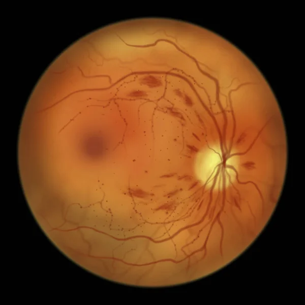
Diabetic Retinopathy Non-proliferative, Illustration Showing Hard Exudates, Cotton Wool Spots, Microaneurysms, Dot Haemorrhages, Flame-shaped And Splinter Retinal Haemorrhages, IRMAs, Venous Beading
Image, 3.07MB, 5000 × 5000 jpg
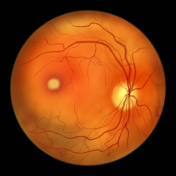
Best Disease. Best Vitelliform Macular Dystrophy, Vitelliform Stage, Classic Egg-yolk Lesion, Scientific Illustration, Ophthalmoscope View
Image, 2.87MB, 5000 × 5000 jpg

Microaneurysms, Microscopic Buldges In The Artery Walls Filled With Blood, 3D Illustration. Found In The Eye Retina In Diabetic Retinopathy, And Also In Brain (Charcot-Bouchard Aneurysms)
Image, 7.58MB, 7200 × 4050 jpg

Retinal Arteriovenous Malformation: Rare Congenital Retinal Vascular Anomalies With Tangled Blood Vessels In The Retina, Illustration Shows Artery-vein Communication Without Intervening Capillaries.
Image, 2.64MB, 5000 × 5000 jpg

Diabetic Macular Edema (DME), Illustration Shows Cystoid Macula Edema And Optic Disk Edema, Abnormal Finding On Fundoscopic Examination Of The Eye Retina In Diabetes Mellitus, Fluorescein Angiography
Image, 2.64MB, 5000 × 5000 jpg


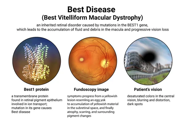
Best Disease. Best Vitelliform Macular Dystrophy, 3D Illustration Showing Best1 Protein, Classic Fundoscopic Egg-yolk Lesion On Retina, And Distorted Vision With Black Spot In A Patient
Image, 7.56MB, 9000 × 6000 jpg

Non-proliferative Diabetic Retinopathy, 3D Illustration Showing Normal Eye Retina And Retina With Hard Exudates, And Cotton Wool Spots
Image, 25.26MB, 11738 × 6603 jpg

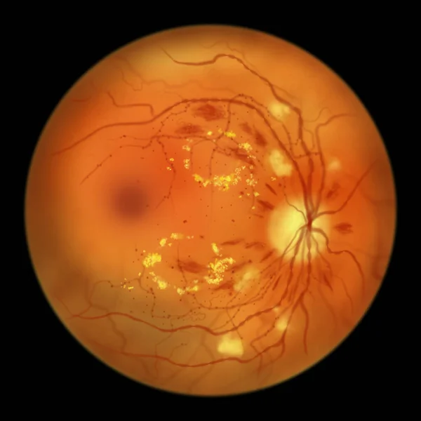
Diabetic Retinopathy, Ophthalmoscope View, Illustration Showing Accumulation Of Fatty Substances Leaked From Blocked Capillaries (yellow Patches), Haemorrhages (red Spots), Microaneurysms
Image, 4.38MB, 5000 × 5000 jpg

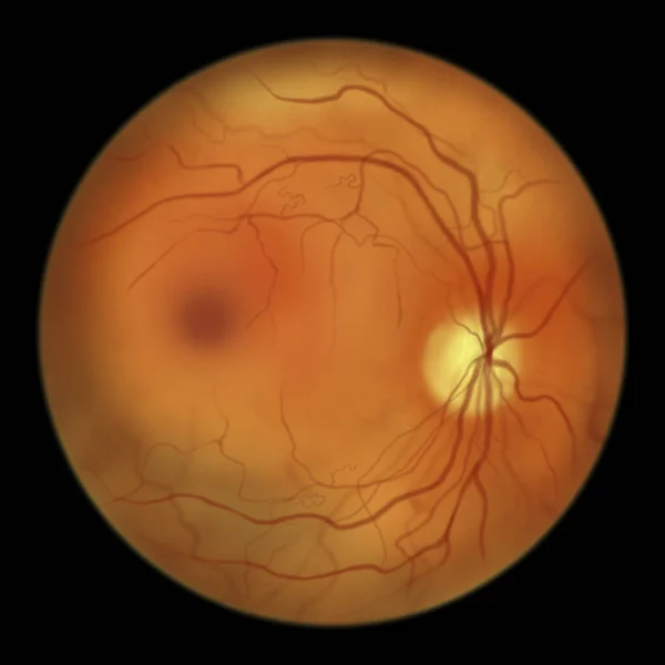
Proliferative Diabetic Retinopathy, Illustration Showing Neovascularization In The Optic Disk And Other Sites. Fundoscopic Examination Of The Eye Retina In Diabetes Mellitus, Fluorescein Angiography
Image, 6.07MB, 11738 × 6603 jpg
A Medical 3D Illustration Showcasing A Healthy, Normal Retina As Observed During Ophthalmoscopy, Displaying Clear Retinal Structures And Vasculature.
Image, 10.3MB, 5352 × 5352 jpg
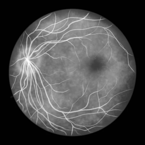
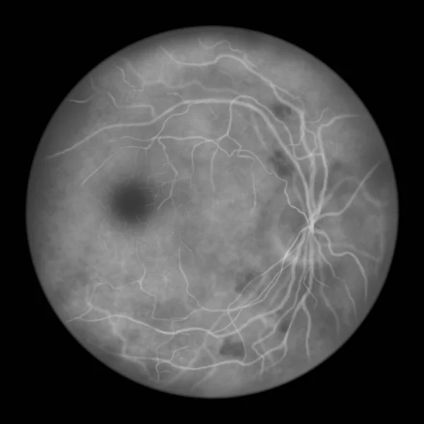
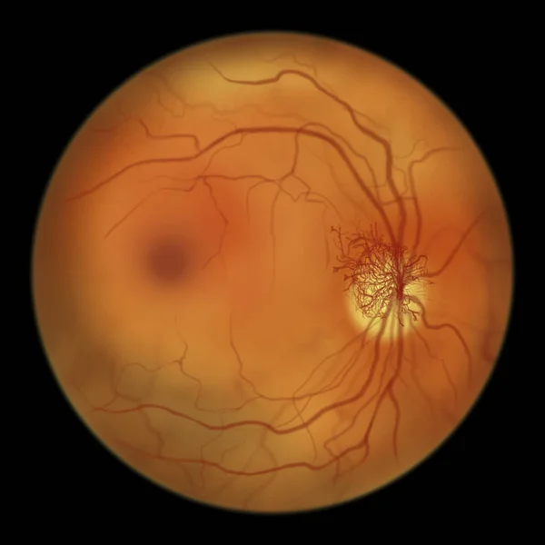
Normal Eye Retina, Scientific Illustration Showing Optic Disk, Blood Vessels, Macula And Fovea, Ophthalmoscope View, Fluorescein Angiography
Image, 2.91MB, 5000 × 5000 jpg
Page 1 >> Next