Home
Images
Vectors
Videos
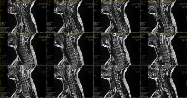
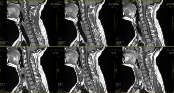
Stock image set of 6 sagittal MRI scans of neck area of caucasian 34 years old male with bilateral paramedial extrusion of the C6-C7 segment with radiculopathy
Published:
Jun.02, 2023 15:46:27
Author:
z1bjkeee
Views:
13
Downloads:
0
File type:
image
/
jpg
File size:
2.52 MB
Orginal size:
3840
x
2055
px
Available sizes:
S
(500 x 268)
M
(1000 x 535)
L
(2000 x 1070)
XL
(3840 x 2055)
EL0
(3840 x 2055)
Level:
bronze
Download Image
Image topics:
Computer
Set
Side
Human
Caucasian
Medicine
Healthcare
Male
Medical
Technology
Hospital
Bone
Neck
Information
Disease
Serious
Pain
Compression
Slices
Suffering
Spine
Plane
Magnetic
Internal
Diagnosis
Examination
Anatomy
X Ray
Imaging
Resonance
Radiology
Xray
Bilateral
Radiography
Mri
Extrusion
Ct
Vertebrae
Compressive
Scans
Hernia
Sagittal
C6
Paresis
Radiculopathy
Left Sided
Paramedial
Similar stock images
Set Of Sagittal MRI Scans Of Neck Area Of Caucasian 34 Years Old Male With Bilateral Paramedial Extrusion Of The C6-C7 Segment With Radiculopathy
3840 × 2019
Set Of Sagittal MRI Scans Of Neck Area Of Caucasian Male With Bilateral Paramedial Extrusion Of The C6-C7 Segment With Radiculopathy
3000 × 2000
Mri Of A Lumbar Disc Herniation-
4000 × 4000
Magnetic Resonance Images Of Cervical Spine Sagittal T1-weighted Images (MRI Cervical Spine) Showing Mild Disc Disease.
5760 × 3240
MRI OF THORACIC SPINE HISTORY: A 53-year-old Woman, Presented Wi
5144 × 3900
MRI Scan
3631 × 2421
Mri Of A Lumbar Disc Herniation-
4000 × 4000
Magnetic Resonance Images Of Lumbar Spine Sagittal T1-weighted Images (MRI Lumbar Spine) Showing Mild Disc Disease.
5760 × 3240