photo free
Home Images Vectors Videos

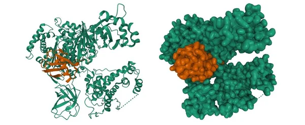
Stock image Structure of coagulation factor IXa, 3D cartoon and Gaussian surface models, chain id color scheme, based on PDB 6mv4, white background
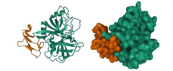
Stock image Structure of coagulation factor IXa, 3D cartoon and Gaussian surface models, chain id color scheme, based on PDB 6mv4, white background
Published: Jul.29, 2021 08:53:26
Author: unnaugan
Views: 10
Downloads: 0
File type: image / jpg
File size: 4.63 MB
Orginal size: 10000 x 4100 px
Available sizes:
S
(500 x 205)
M
(1000 x 410)
L
(2000 x 820)
XL
(10000 x 4100)
EL0
(10000 x 4100)
Level: beginner
Download Image
Image topics:

Similar stock images

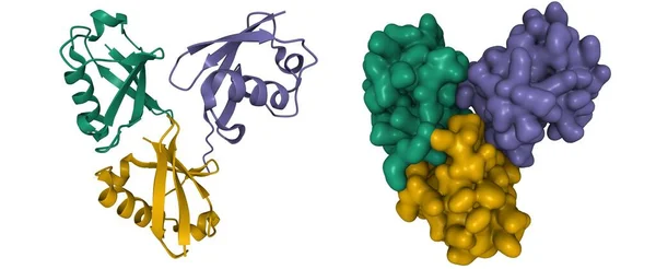
stock image Crystal Structure of Branched K11-K48-Linked Tri-Ubiquitin, 3D cartoon and Gaussian surface models, based on PDB 9oq1, white background

Crystal Structure Of Branched K11-K48-Linked Tri-Ubiquitin, 3D Cartoon And Gaussian Surface Models, Based On PDB 9oq1, White Background

10000 × 4100
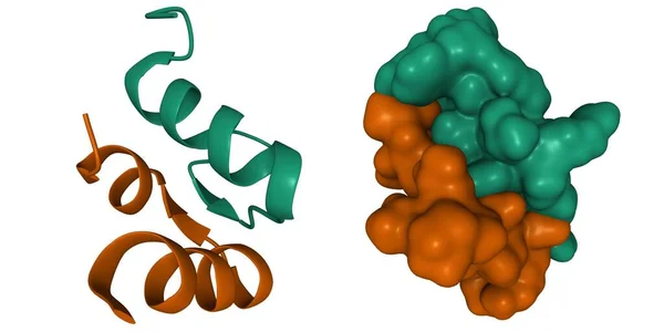
stock image Structure of human hormone relaxin-3 heterodimer, 3D cartoon and Gaussian surface models, white background

Structure Of Human Hormone Relaxin-3 Heterodimer, 3D Cartoon And Gaussian Surface Models, White Background

8000 × 4000
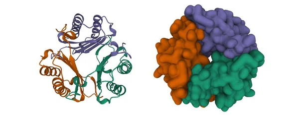
stock image Structure of human Macrophage Migration Inhibitory Factor (Glycosylation-Inhibiting Factor) homotrimer. 3D cartoon and Gaussian surface models, chain id color scheme, PDB 1gif, white background

Structure Of Human Macrophage Migration Inhibitory Factor (Glycosylation-Inhibiting Factor) Homotrimer. 3D Cartoon And Gaussian Surface Models, Chain Id Color Scheme, PDB 1gif, White Background

10000 × 4000
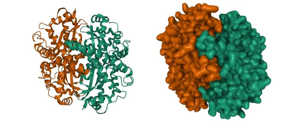
stock image Structure of native human manganese superoxide dismutase, 3D cartoon and Gaussian surface models, chain id color scheme, based on PDB 5vf9, white background

Structure Of Native Human Manganese Superoxide Dismutase, 3D Cartoon And Gaussian Surface Models, Chain Id Color Scheme, Based On PDB 5vf9, White Background

10000 × 4100
stock image Structure of human ubiquitin activating enzyme E1 (Uba1, green) in complex with ubiquitin (brown), 3D cartoon and Gaussian surface model, based on PDB 6dc6, white background

Structure Of Human Ubiquitin Activating Enzyme E1 (Uba1, Green) In Complex With Ubiquitin (brown), 3D Cartoon And Gaussian Surface Model, Based On PDB 6dc6, White Background

10000 × 4100
stock image Crystal structure of human glutathione transferase (GST) A1-1 in complex with glutathione, 3D cartoon and Gaussian surface models, chain id color scheme, based on PDB 1pkw, white background

Crystal Structure Of Human Glutathione Transferase (GST) A1-1 In Complex With Glutathione, 3D Cartoon And Gaussian Surface Models, Chain Id Color Scheme, Based On PDB 1pkw, White Background

10000 × 4000
stock image Structure of human hormone insulin-like peptide-3 heterodimer, 3D cartoon and Gaussian surface models, white background

Structure Of Human Hormone Insulin-like Peptide-3 Heterodimer, 3D Cartoon And Gaussian Surface Models, White Background

10000 × 4000
stock image Crystal structure of human epidermal growth factor dimer. 3D cartoon and Gaussian surface models, chain id color scheme, PDB 1jl9, white background

Crystal Structure Of Human Epidermal Growth Factor Dimer. 3D Cartoon And Gaussian Surface Models, Chain Id Color Scheme, PDB 1jl9, White Background

8000 × 4000