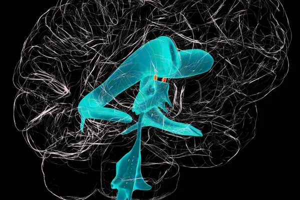
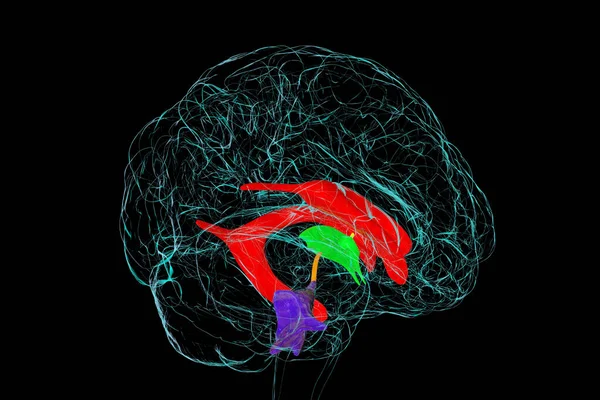
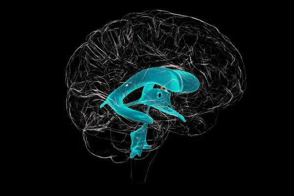
Stock image The interventricular foramen, the foramen of Monro, a passage that connects the brain lateral ventricles, allowing cerebrospinal fluid to flow between them and the third ventricle, 3D illustration.
Published: Jul.07, 2023 08:30:29
Author: katerynakon
Views: 8
Downloads: 1
File type: image / jpg
File size: 5.3 MB
Orginal size: 6000 x 4000 px
Available sizes:
Level: silver