
Stock image Foramen

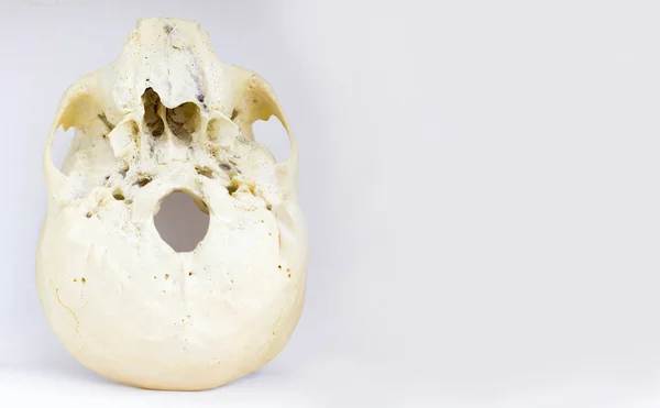
Bottom View Of Base Of The Human Skull Showing Maxilla And Foramen Magnum For Anatomy In Isolated White Background.
Image, 4.24MB, 5588 × 3456 jpg
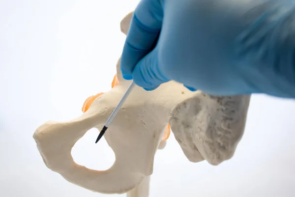
Doctor, Scientist, Specialist In Anatomy Indicates Pointer Of Obturator Foramen Where Canalis Obturatorius, Involving Obturator Artery, Vein And Nerve And Connects Pelvis To Thigh, Place Of Hernia
Image, 4.59MB, 6000 × 4000 jpg

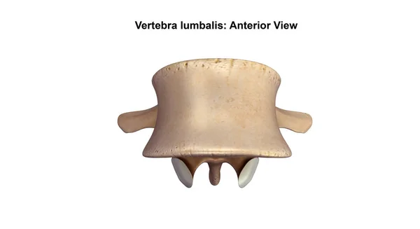
Medical Education Chart Of Biology Human Skull Diagram. Vector. Front Aspect White Background Basic Medical Education
Vector, 1.21MB, 5333 × 4000 eps

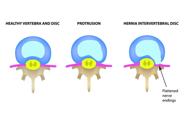
Protrusion Of The Intervertebral Disc. Hernia. Vector Illustration On Isolated Background.
Vector, 1.41MB, 5000 × 3245 eps

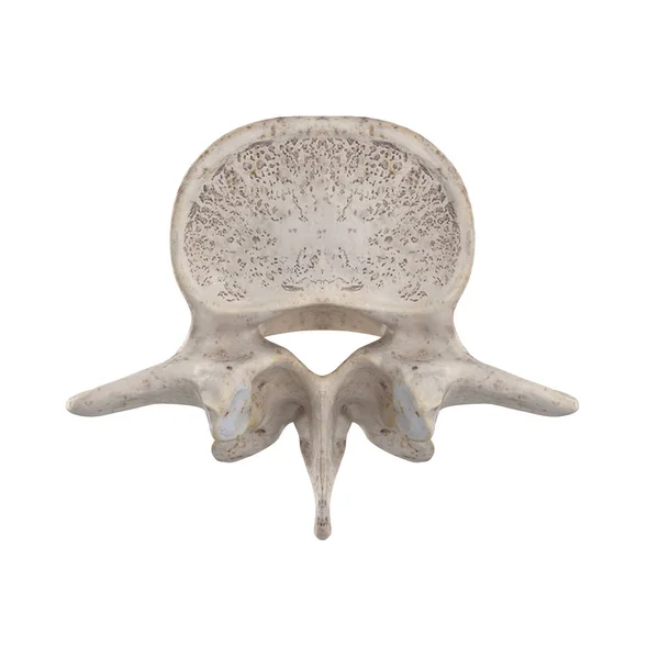
Central Lateral Stenosis, Second Lumbar Vertebra, Nervous System, Spinal Cord, Lumbar Spine, Nerve Root, Advanced Uncovertebral Arthrosis Of The Lumbar Vertebra, Degenerative Changes Vertebra, Osteophytes, Spondylophytes, Osteoarthritis Of The Joints
Image, 7.58MB, 5906 × 5178 jpg
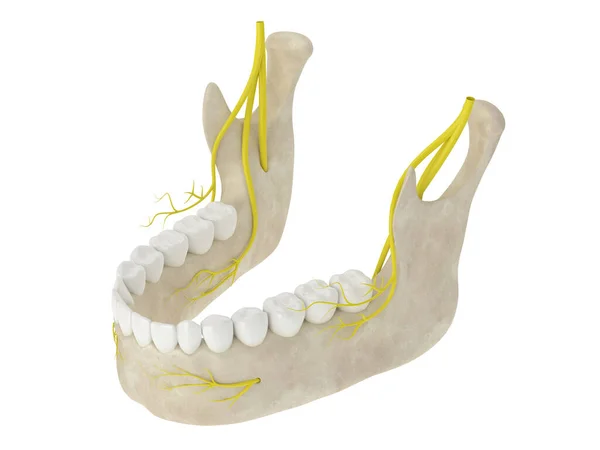
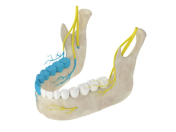
3d Render Of Mandibular Arch With Nerves Isolated Over White Background
Image, 2.41MB, 5000 × 3750 jpg
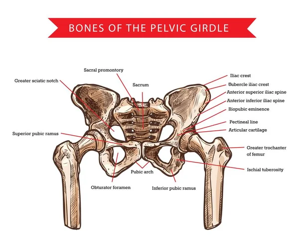
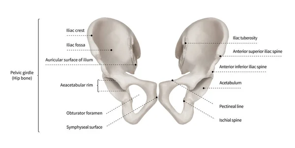
Pelvis Bones Of Pelvic Girdle, Vector Sketch Of Human Anatomy And Medicine. Bones And Joints Structure Of Skeleton Hips, Sacrum, Femur And Coccyx, Sacral Promontory, Pubic Arch And Iliac Spine
Vector, 2.56MB, 5957 × 5036 eps
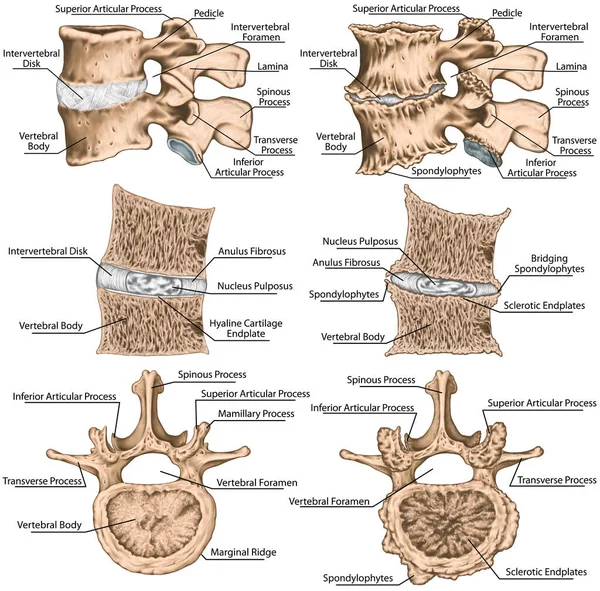
Didactic Board, Spondylophytes Involving A Spinal Motion Segment, Advanced Uncovertebral Arthrosis, Third And Fourth Lumbar Vertebrae, Degenerative Changes Vertebra, Lumbar Vertebra, Lumbar Spine, Vertebral Bone
Image, 9.14MB, 5906 × 5817 jpg

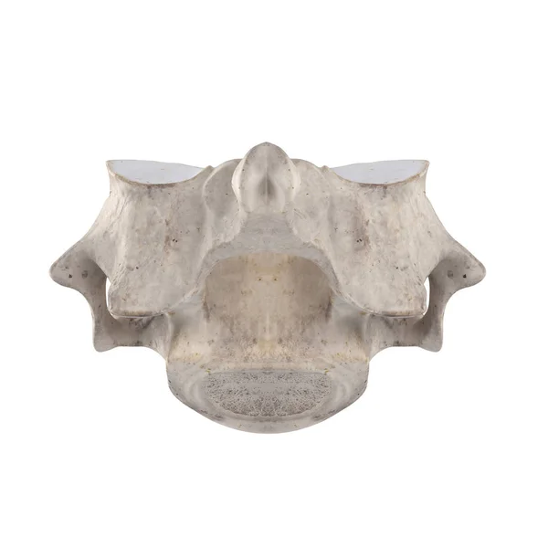
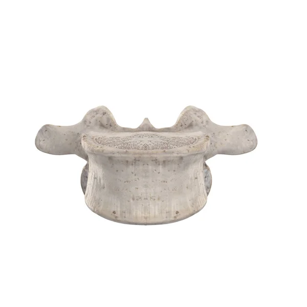
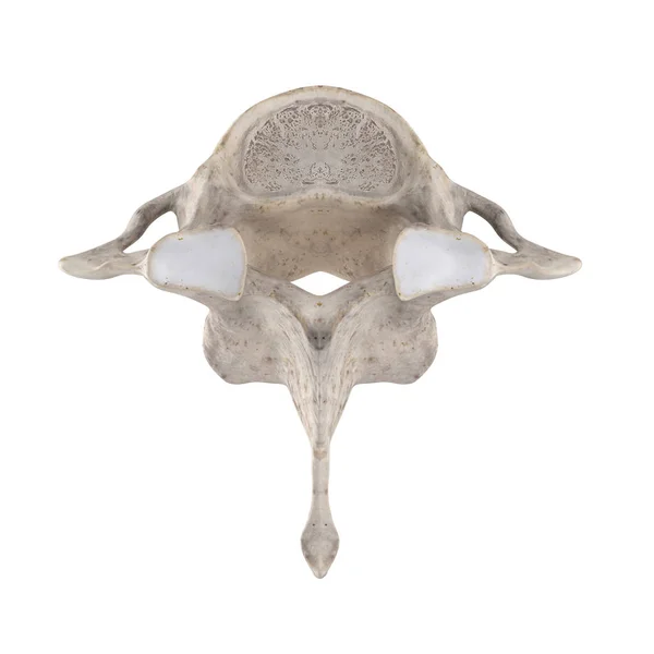
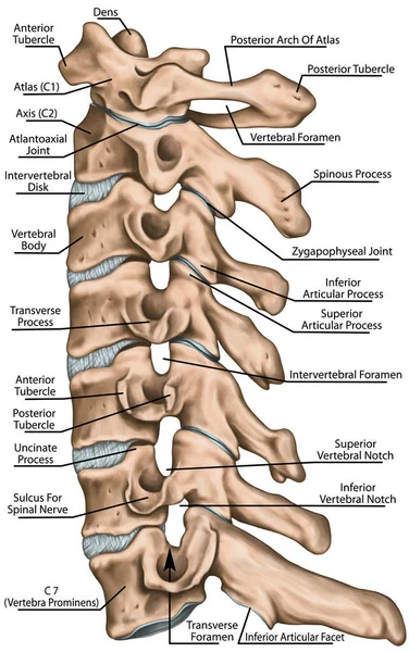
The Ligaments Of The Median Atlantoaxial Joint. Atlas And Axis Ligaments. Cervical Spine, Vertebral Morphology, First And Second Cervical Vertebra, Cervical Vertebrae, Atlas, Axis, Atlantoaxial Joint, Superior View
Image, 4.03MB, 5906 × 4229 jpg
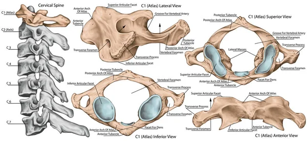
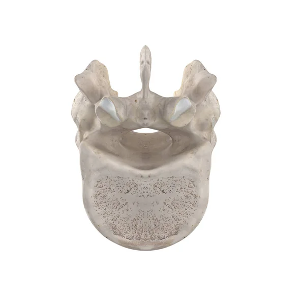
Didactic Board, Cervical Spine, Vertebral Morphology, First Cervical Vertebra, Atlas, Cervical Vertebrae, Anterior, Lateral, Superior And Inferior View
Image, 4.48MB, 5906 × 2715 jpg

3d Render Of Mandibular Arch Showing Blocked Inferior Alveolar Nerve Area. Types Of Dental Anesthesia Concept.
Image, 2.37MB, 5000 × 3750 jpg
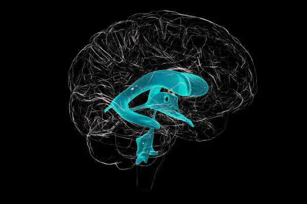
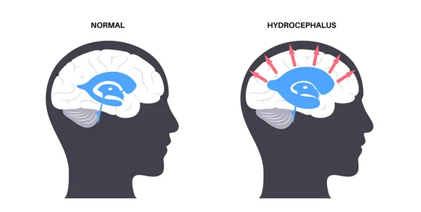
The Interventricular Foramen, The Foramen Of Monro, A Passage That Connects The Brain Lateral Ventricles, Allowing Cerebrospinal Fluid To Flow Between Them And The Third Ventricle, 3D Illustration.
Image, 5.3MB, 6000 × 4000 jpg
Skull - High Quality Vector Logo - Vector Illustration Ideal For T-shirt Graphic
Vector, 0.09MB, 2400 × 2928 eps
Infographic Diagram Of Human Hip Bone Or Pelvic Girdle Anatomy System Anterior View- 3D- Human Anatomy- Medical Diagram- Educational And Human Body Concept- Isolated On White Background
Image, 8.54MB, 15000 × 7536 jpg

Spondylophytes Involving A Spinal Motion Segment, Third And Fourth Lumbar Vertebrae, Advanced Uncovertebral Arthrosis, Degenerative Changes Vertebra, Osteophytes, Osteoarthritis Of The Joints, Lumbar Vertebra, Lumbar Spine, Vertebral Bone
Image, 5.09MB, 5906 × 3518 jpg
Skull - High Quality Vector Logo - Vector Illustration Ideal For T-shirt Graphic
Vector, 0.01MB, 3713 × 3713 eps

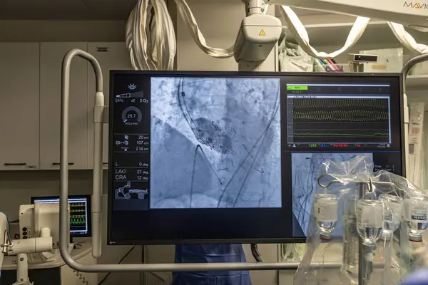
Istanbul, Turkey February 29, 2024; X-ray For Valvular Replacement With Melody Valve. View Of Prosthetic Heart Valve Dysfunction In Patient Who Has Mitral Valve Replacement With Mechanical Valve.
Image, 9.9MB, 5000 × 3335 jpg
Istanbul, Turkey February 29, 2024; X-ray For Valvular Replacement With Melody Valve. View Of Prosthetic Heart Valve Dysfunction In Patient Who Has Mitral Valve Replacement With Mechanical Valve.
Image, 9.91MB, 5000 × 3335 jpg

Istanbul, Turkey February 29, 2024; X-ray For Valvular Replacement With Melody Valve. View Of Prosthetic Heart Valve Dysfunction In Patient Who Has Mitral Valve Replacement With Mechanical Valve.
Image, 10.13MB, 5000 × 3335 jpg

Istanbul, Turkey February 29, 2024; X-ray For Valvular Replacement With Melody Valve. View Of Prosthetic Heart Valve Dysfunction In Patient Who Has Mitral Valve Replacement With Mechanical Valve.
Image, 10.02MB, 5000 × 3335 jpg
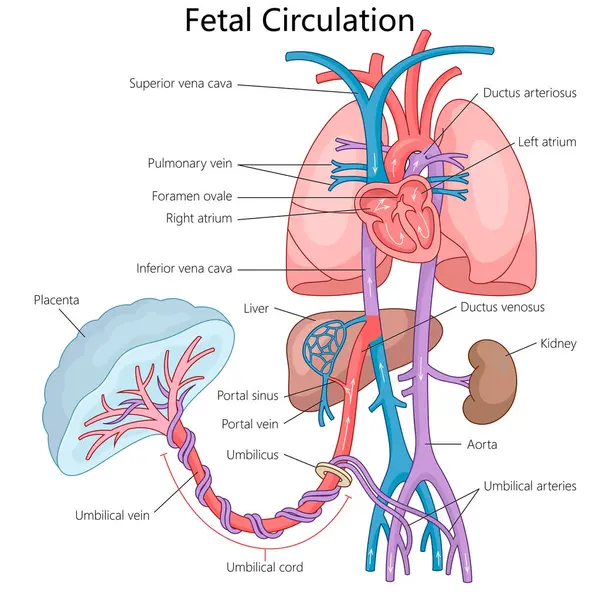
Fetal Circulation System, Blood Flow From The Placenta Through The Umbilical Cord To Heart And Other Organs Diagram Hand Drawn Schematic Raster Illustration. Medical Science Educational Illustration
Image, 5.64MB, 6000 × 6000 jpg
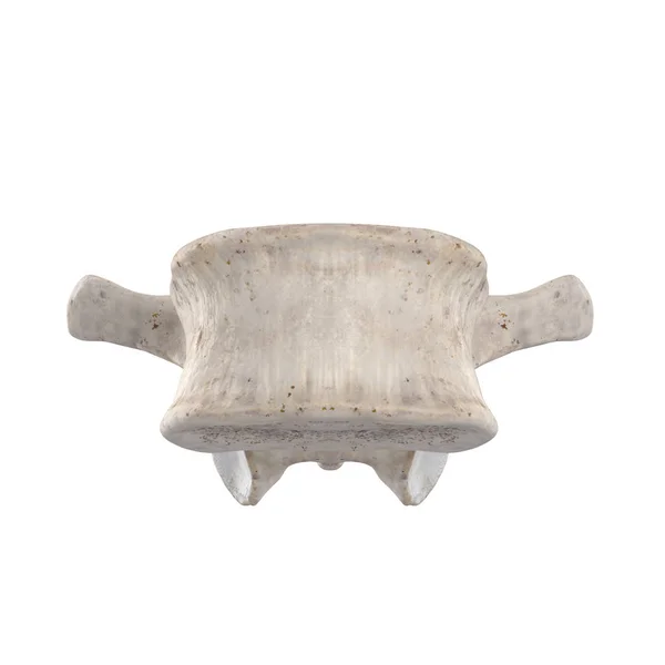


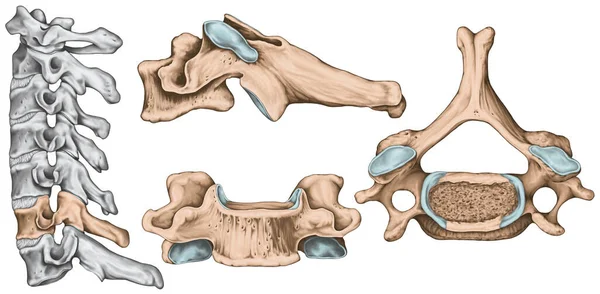
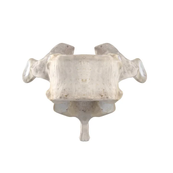
Didactic Board, Cervical Spine, Common Vertebral Morphology, Sixth Cervical Vertebra, Cervical Vertebrae, Anterior, Lateral And Superior View
Image, 3.91MB, 5906 × 2902 jpg

Human Sacrum Bone Structure Diagram, Anatomical Vector Illustration Labeled Scheme With Bone Sections.
Vector, 5.28MB, 4655 × 3766 eps


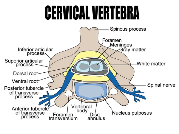
Labeled Vertebra Cross Section Of Human Body Anatomy Infographic Diagram Including All Parts Cord Of Grey And White Matter Spinal Nerve Vertebral Body Foramen And Spinous Process For Medical Science Education And Healthcare
Vector, 0.64MB, 2619 × 1593 eps
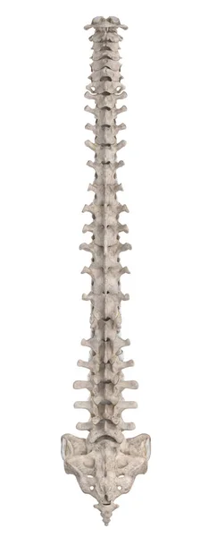
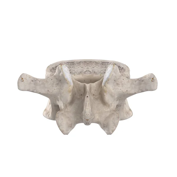
Cervical Spine Structure, Vertebral Bones, Cervical Bones, Anatomy Of Human Bone System, Human Skeletal System, Vertebral Morphology, Lateral View
Image, 4.52MB, 3304 × 5268 jpg
Page 1 >> Next