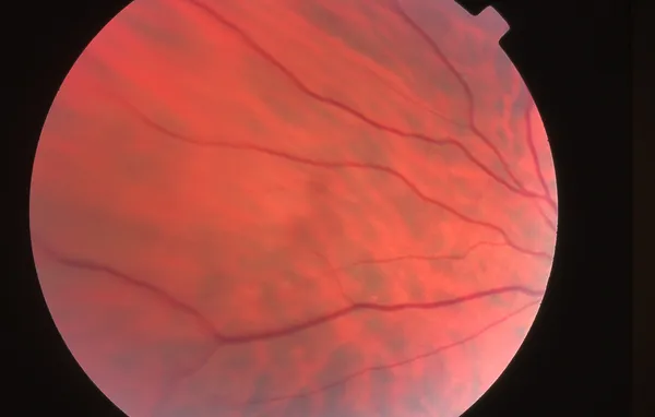
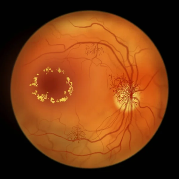
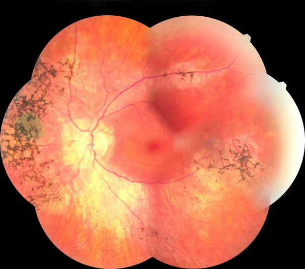
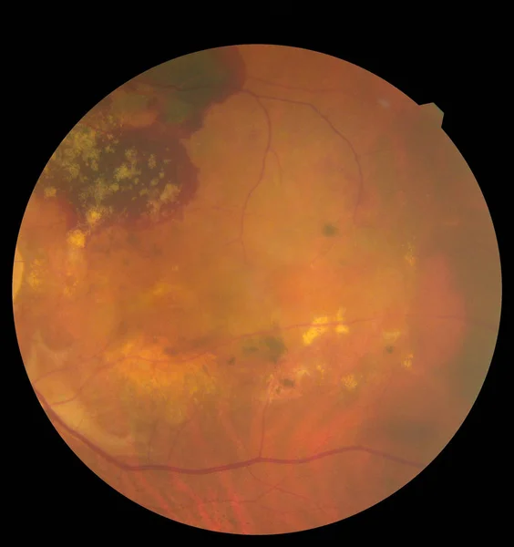
Stock image View inside human eye disorders showing retina, optic nerve and macula. Retinal picture ,Medical photo tractional eye screen retinal detachment of diabetes. Eye treatment concept.
Published: Jan.28, 2020 16:13:37
Author: ternavskaia.o@gmail.com
Views: 32
Downloads: 5
File type: image / jpg
File size: 2.62 MB
Orginal size: 3000 x 3186 px
Available sizes:
Level: bronze