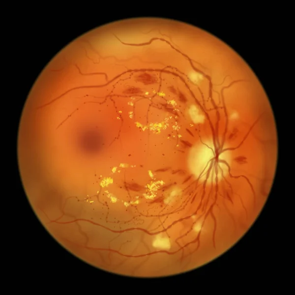
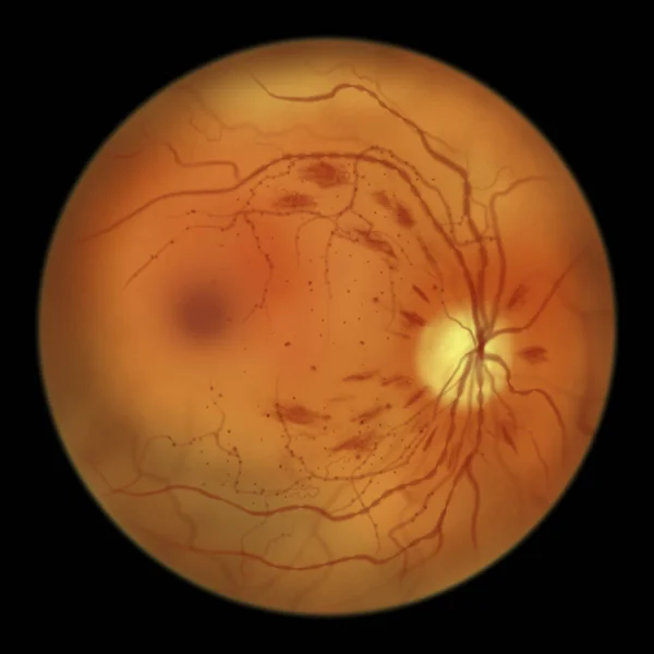
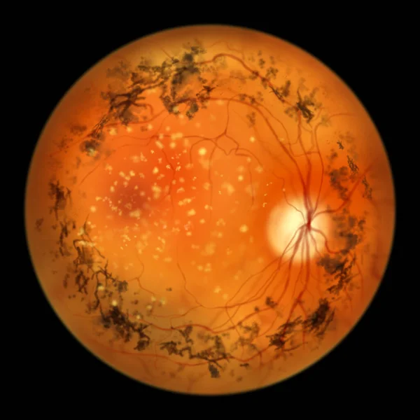
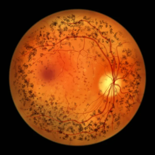
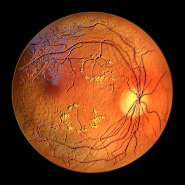
Stock image Diabetic retinopathy, ophthalmoscope view, illustration showing accumulation of fatty substances leaked from blocked capillaries (yellow patches), haemorrhages (red spots), microaneurysms

Published: Apr.07, 2022 10:15:44
Author: katerynakon
Views: 11
Downloads: 5
File type: image / jpg
File size: 4.38 MB
Orginal size: 5000 x 5000 px
Available sizes:
Level: silver