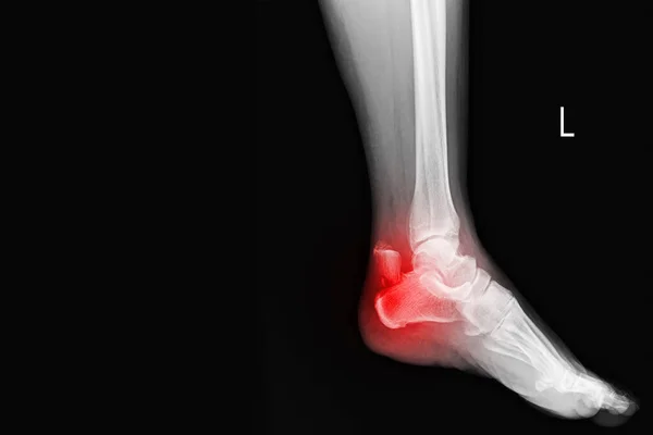
Stock image X-ray Foot and Ankle Normal joint and osteochondroma of distal tibia cause pressure effect to fibula.Medical image concept.

Published: Jul.19, 2021 10:32:06
Author: Richmanphoto
Views: 6
Downloads: 0
File type: image / jpg
File size: 4.41 MB
Orginal size: 6000 x 4000 px
Available sizes:
Level: bronze
Similar stock images

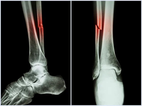
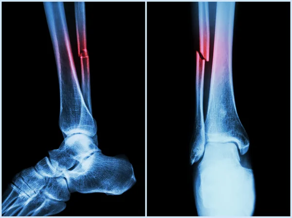
Fracture Shaft Of Fibula Bone ( Leg Bone ) . X-ray Of Leg ( 2 Position : Side And Front View )
7032 × 5264
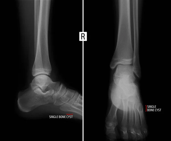
X-ray Of The Ankle Joint. Shows The Bony Cyst Of The Fifth Finger Of The Right Foot. Marker.
2976 × 2454
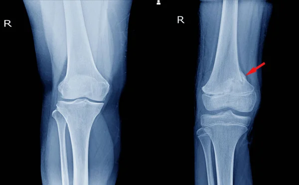
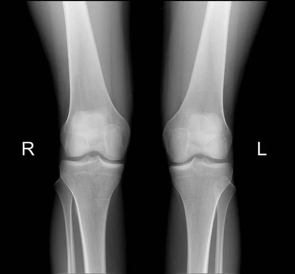
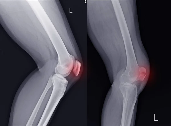
X-ray Knee AP Views Showing Normal Knee Joint And Fracture Distal Femur On Red Arrow Point.
5440 × 3368