Home
Images
Vectors
Videos
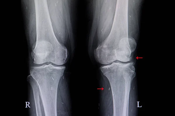
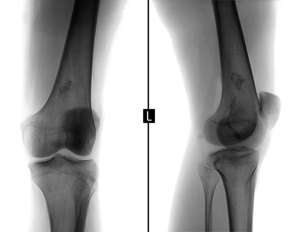
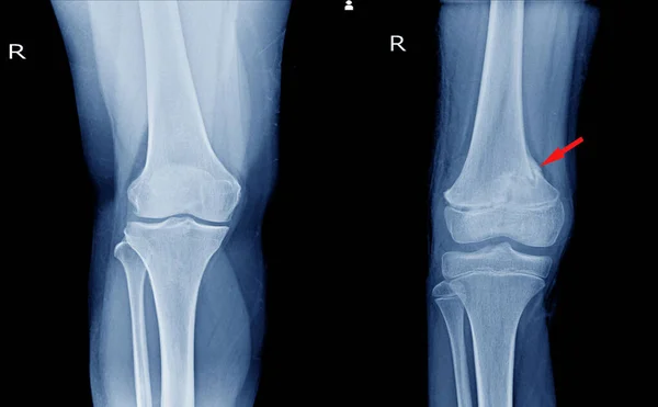
Stock image X-ray Knee AP views Showing Normal knee joint and fracture distal femur on red arrow point.
Published:
Mar.30, 2020 06:10:46
Author:
Richmanphoto
Views:
184
Downloads:
0
File type:
image
/
jpg
File size:
5.12 MB
Orginal size:
5440
x
3368
px
Available sizes:
S
(500 x 310)
M
(1000 x 619)
L
(2000 x 1238)
XL
(5440 x 3368)
EL0
(5440 x 3368)
Level:
bronze
Download Image
Image topics:
Image
Human
People
Health
Medicine
Illness
Medical
Care
Clinical
Doctor
Hospital
Bone
Patient
Therapy
Treatment
Broken
Film
Leg
Pain
Tissue
Knee
Thigh
Joint
Skeleton
Fracture
Cast
Diagnosis
Examination
Anatomy
X Ray
Surgery
Surgical
Trauma
Accident
Injury
Scan
Operation
Radiology
Xray
Swelling
Skeletal
Orthopedic
Tibia
Femur
Fibula
Patella
Kneecap
Shinbone
Distal
Thighbone
Similar stock images
Xray Image Of Knees With Degenenative Osteoarthritis And Calcified Soft Tissue Cysticercosis. Chronic Knee Pain.
6240 × 4160
Knee Joint X-ray Showing Fracture Distal Femur (thigh Bone)on Red Mark.and Black Background. Medical Healthcare Concept.
6000 × 4000
X-ray Foot And Ankle Normal Joint And Osteochondroma Of Distal Tibia Cause Pressure Effect To Fibula.Medical Image Concept.
6000 × 4000
X-ray Left Knee Lateral Showing Kneecap Normal And Fracture Red Color Marker.
3800 × 2800
X-ray Knee Radiograph Show State Of Injury
3989 × 2422
Osteoarthritis (OA) Knee . Film X-ray APand Lateral View Of Knee Show Narrow Joint Space, Osteophyte ( Spur ), Subchondral Sclerosis, Knee Joint Inflammation.
5493 × 4500
X-ray Of The Left Knee Joint. Ewing Sarcoma, Lymphoma, Myeloma Thigh Bone. Negative.
3250 × 2539
X-ray Image Of Fracture Leg (tibia )with Implant Plate And Scre
5184 × 3456