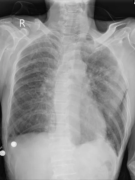
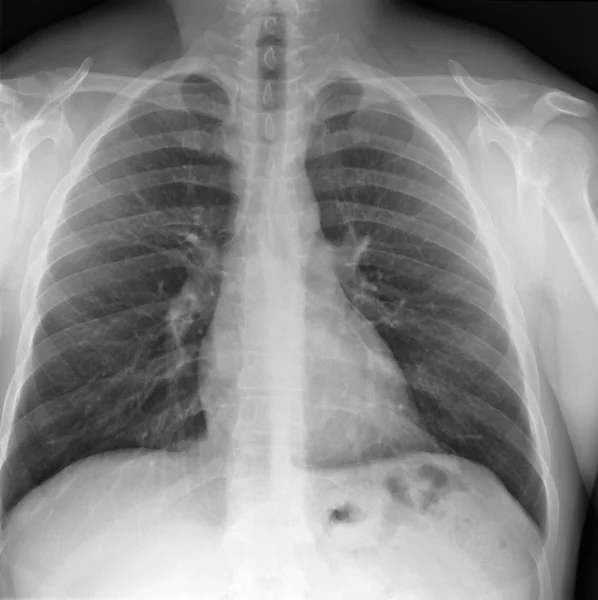
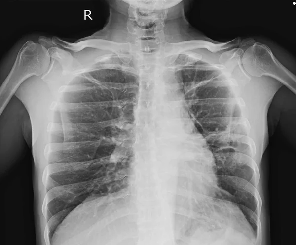
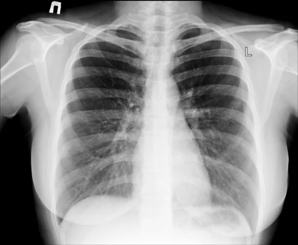
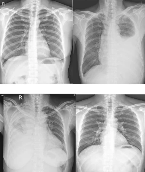
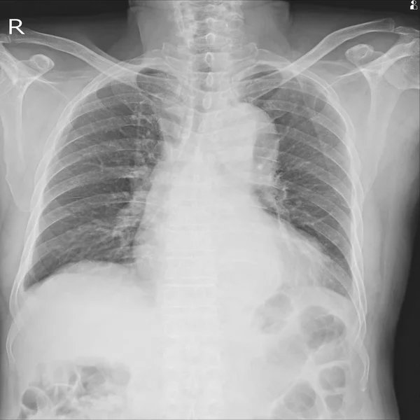
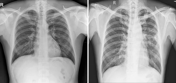
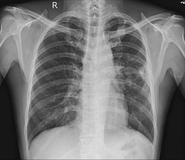
Stock image X-ray image of the chest showing the internal anatomy of the rib
Published: Jul.10, 2017 08:43:44
Author: gubernat
Views: 1211
Downloads: 7
File type: image / jpg
File size: 27.51 MB
Orginal size: 12000 x 9830 px
Available sizes:
Level: beginner