Home
Images
Vectors
Videos
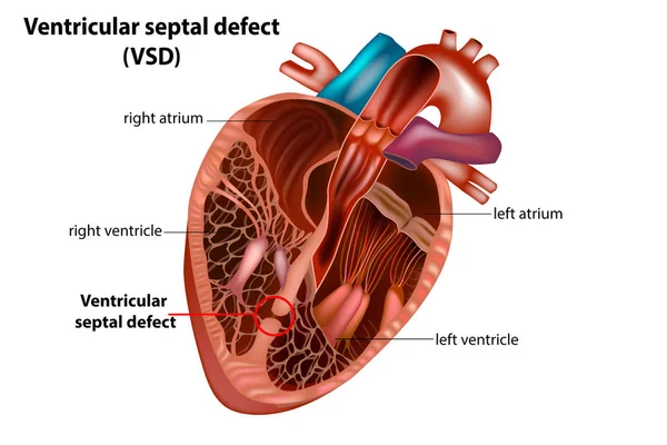
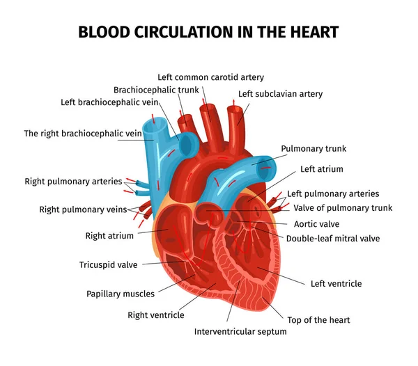
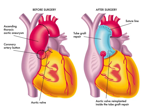
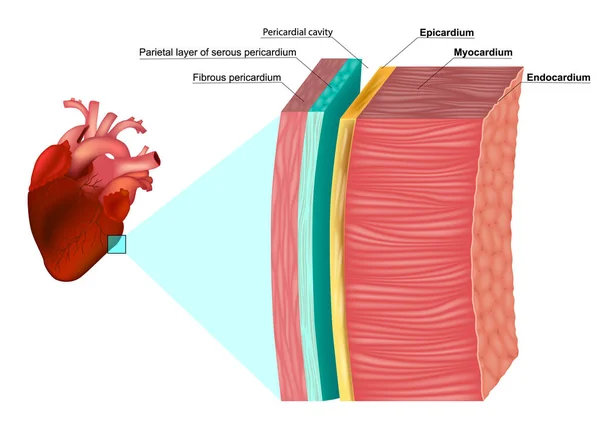
Stock vector The Layers of the Heart Wall Anatomy. Myocardium, Epicardium, Endocardium and Pericardium. Heart wal structure diagram
Published:
Mar.01, 2023 10:42:19
Author:
Sakurra
Views:
46
Downloads:
3
File type:
vector
/
eps
File size:
8.15 MB
Orginal size:
6300
x
4500
px
Available sizes:
S
(500 x 357)
M
(1000 x 714)
L
(2000 x 1429)
XL
(6300 x 4500)
VECT
(6300 x 4500)
EL0
(6300 x 4500)
Level:
bronze
Download Vector
Vector topics:
Vector
Illustration
Closeup
Human
Health
Medicine
Medical
Structure
Wall
Cardiology
Cross
Heart
Body
Education
Biology
Science
Section
System
Organ
Muscle
Layer
Membrane
Location
Scheme
Layers
Educational
Anatomy
Diagram
Cardiovascular
Cavity
Circulation
Cardiac
Fibrous
Anatomical
Vecto
Serous
Aorta
Parietal
Coronary
Visceral
Myocardium
Infographic
Pericardium
Endocardium
Epicardium
Pericardial
Similar stock vectors
Ventricular Septal Defect (VSD).Congenital Heart Defect
5001 × 3334
Heart Blood Flow Composition
5127 × 4500
Thoracic Aortic Aneurysm Surgery
5333 × 4000
Stent With Balloon Angioplasty
5617 × 3918
Realistic Detailed 3d Stomach Anatomy Internal Organ. Vector
5222 × 5222
Late-stage Scarring Of Liver Illustration
5905 × 3763
Diagram Showing Cirrhosis Of The Liver
6808 × 3231
A Comparison Of Peripheral Artery Disease Illustration
6444 × 3414