
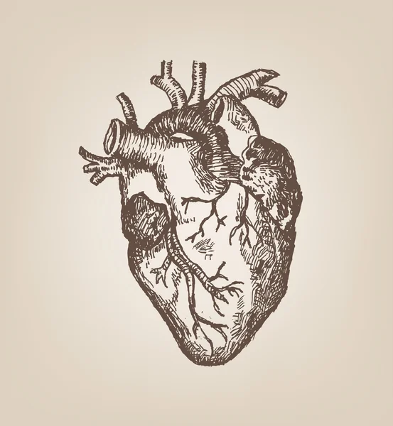
Stock image Epicardium
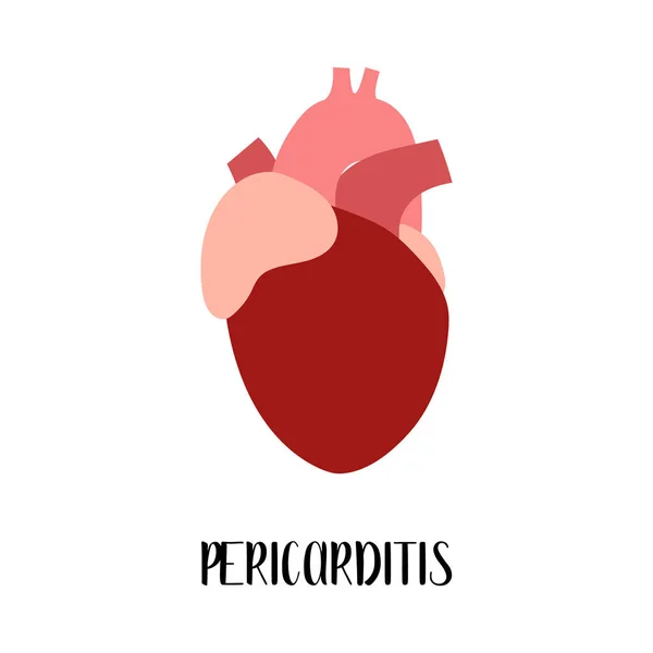
Pericarditis. Heart, Cardiovascular Diseases. Cardiology. Vector Flat Illustration. For Flyer, Medical Brochure, Banner, Landing Page, Web
Vector, 5.32MB, 4000 × 4000 eps

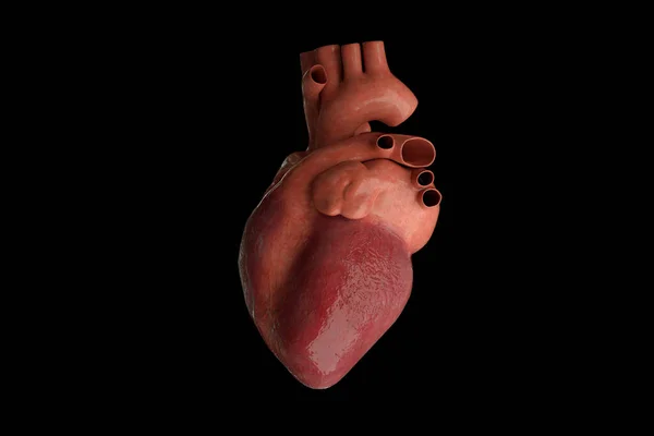
Healthy Heart, Cardiovascular System. Cardiology. Vector Flat Illustration. For Flyer, Medical Brochure, Banner, Landing Page, Web
Vector, 5.32MB, 4000 × 4000 eps

Cardiac Muscle Tissue Structure. Myocardium Anatomical Poster. Cardiomyocytes Cells. Walls Of The Heart In The Human Body, Relaxation And Contraction Of Muscle Fibers Flat Vector Medical Illustration.
Vector, 0.55MB, 7767 × 4345 eps

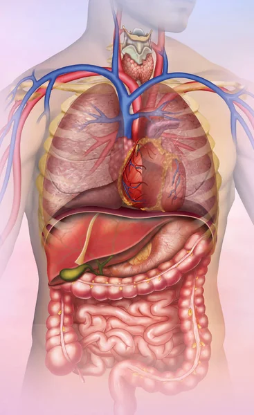
In Humans, Purkinje Fibers Are Not Distributed Throughout The Entire Ventricular Wall, But Rather In The Superficial Myocardium Beneath The Endocardium And Do Not Reach The Epicardium.
Image, 7.05MB, 10000 × 10375 jpg

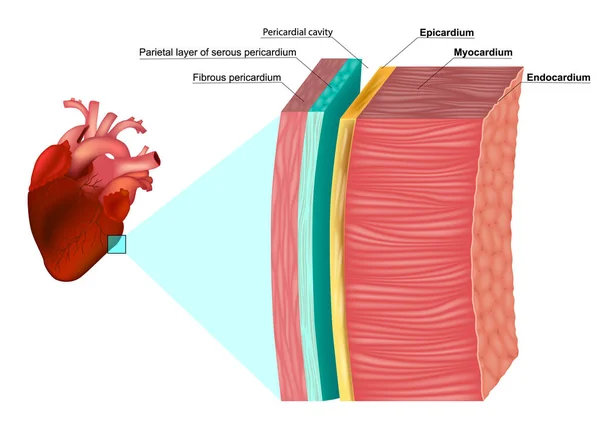
Layers Of Heart Wall. Pericardium Structure. Anatomy Of Pericardial Sac. Vector Illustration
Vector, 13.58MB, 4444 × 4010 eps
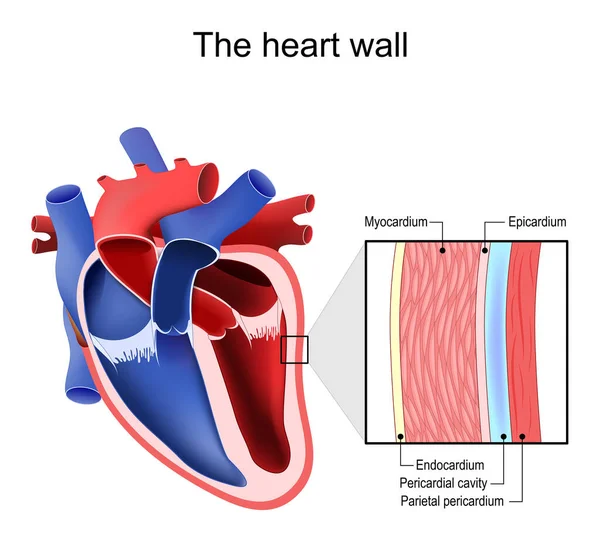
The Layers Of The Heart Wall Anatomy. Myocardium, Epicardium, Endocardium And Pericardium. Heart Wal Structure Diagram
Vector, 8.15MB, 6300 × 4500 eps

Skeleton With Muscles, Joints, Cartilages And Ligaments In The Human Body. Muscle Fibers And Connective Tissue Sheaths In The Female Silhouette. Musculoskeletal Anatomy Vector Medical Illustration
Vector, 1.07MB, 5264 × 3551 eps

Cardiac Muscle Tissue Structure. Myocardium Anatomical Poster. Cardiomyocytes Cells. Walls Of The Heart In The Human Body, Relaxation And Contraction Of Muscle Fibers Flat Vector Medical Illustration.
Vector, 0.55MB, 7683 × 3701 eps

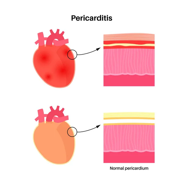
Pericarditis Anatomical Poster. Heart Wall Inflammation. Chest Pain Symptom. Inflamed Internal Organs Concept. Viral Infection In The Human Body. Cardiovascular System Medical Flat Vector Illustration
Vector, 0.51MB, 4168 × 4167 eps

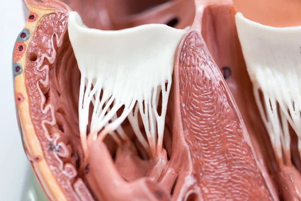
During The Onset Of Variant Angina Pectoris, ECG Is Divided Into Non Fusion Wave, Partial Fusion Wave And Complete Fusion Wave According To The Fusion Degree Of QRS Wave, ST Segment And T Wave.
Image, 7.94MB, 10000 × 6537 jpg

Pericardial Effusion Poster. Fluid In The Space Around The Heart, Cardiac Tamponade Cause. Inflamed Internal Organs, Infection In The Human Body. Cardiovascular System Medical Vector Illustration
Vector, 0.36MB, 4764 × 3646 eps
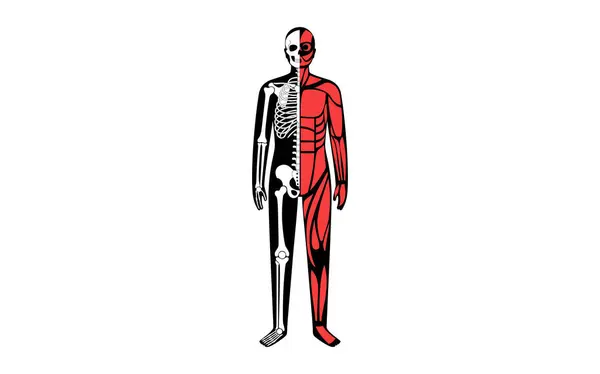
Skeleton With Muscles, Joints, Cartilages And Ligaments In The Human Body. Muscle Fibers And Connective Tissue Sheaths In The Male Silhouette. Musculoskeletal Anatomy Flat Vector Medical Illustration
Vector, 0.53MB, 5694 × 3551 eps
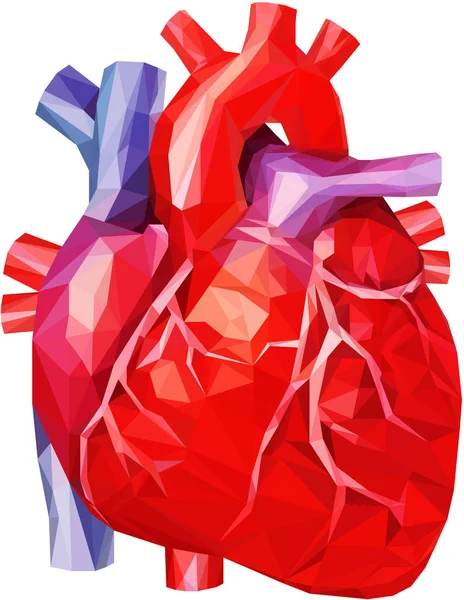
Realistic Human Heart In Low Poly With Veins And Aorta In Red, Purple And Blue
Vector, 1.2MB, 4000 × 5166 eps

Cardiac Muscle Tissue Structure. Myocardium Anatomical Poster. Cardiomyocytes Cells. Walls Of The Heart In The Human Body, Relaxation And Contraction Of Muscle Fibers Flat Vector Medical Illustration.
Vector, 0.5MB, 6666 × 3532 eps

Endocarditis. Heart, Cardiovascular Disease. Cardiology. Vector Flat Illustration. For Flyer, Medical Brochure, Banner, Landing Page, Web
Vector, 5.41MB, 4000 × 4000 eps


Pericardial Effusion Poster. Fluid In The Space Around The Heart, Cardiac Tamponade Cause. Inflamed Internal Organs, Infection In The Human Body. Cardiovascular System Medical Vector Illustration
Vector, 0.35MB, 5164 × 3365 eps

Cardiac Muscle Tissue Structure. Myocardium Anatomical Poster. Cardiomyocytes Cells. Walls Of The Heart In The Human Body, Relaxation And Contraction Of Muscle Fibers Flat Vector Medical Illustration.
Vector, 0.49MB, 7682 × 3701 eps


Cardiac Muscle Tissue Structure. Myocardium Anatomical Poster. Cardiomyocytes Cells. Walls Of The Heart In The Human Body, Relaxation And Contraction Of Muscle Fibers Flat Vector Medical Illustration.
Vector, 0.51MB, 7683 × 3701 eps
Page 1 >> Next