
Stock image Atri page 2

ECG In Atrial Flutter, An Abnormal Heart Rhythm Characterized By Rapid, Regular Contractions Of The Atria. 3D Illustration Displaying Characteristic Sawtooth P-waves And Irregular Ventricular Rhythm.
Image, 9.45MB, 9000 × 6000 jpg

This Photo Shows The Pink Mucinous Matrix Of Atrial Myxoma, Myxoma Cells Arranged In Nests And Cords, And Local Bleeding.Magnify 1000x.
Image, 23.48MB, 4640 × 6267 jpg
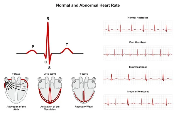
Normal And Abnormal Heart Rate Infographic Diagram Including Activation Of Atria Ventricle And Recovery Wave Also Chart Of Normal Fast Slow Irregular Heartbeats For Medical Science Education And Health Care
Vector, 1.29MB, 2530 × 1642 eps


Peripheral Arterial Occlusive Disease Is The Narrowing Or Blockage Of An Artery In The Legs, Usually Due To Atherosclerosis. Medical Illustration
Image, 1.56MB, 5000 × 3500 jpg


Doctor's Hands In Blue Gloves Shows The Word Endocarditis. Medical Concept.
Image, 2.84MB, 3393 × 1986 jpg
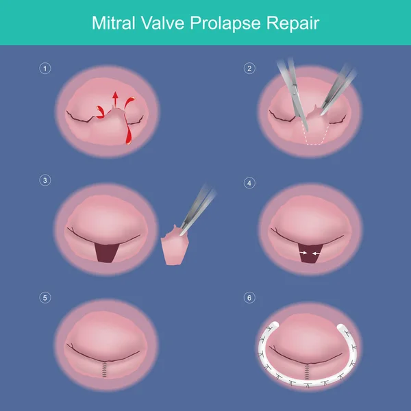
Mitral Valve Prolapse Repair. The Method Repair Heart Valve By Surgery Removed Damaged Or Abnormal Leaked Segment And Used Synthetic Material Ring (medical Parts) For Tissue Strength.
Vector, 28MB, 4000 × 4000 eps

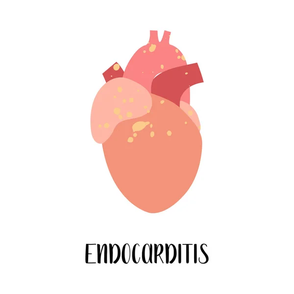
Endocarditis. Heart, Cardiovascular Disease. Cardiology. Vector Flat Illustration. For Flyer, Medical Brochure, Banner, Landing Page, Web
Vector, 5.41MB, 4000 × 4000 eps

Electrocardiogram Displaying A Junctional Rhythm, Which Occurs When The Electrical Signals In The Heart Originate From The Atrioventricular Junction Instead Of The Sinoatrial Node, 3D Illustration.
Image, 7.29MB, 9000 × 6000 jpg


A 67-year-old Man Presents With Heart Palpitations, Numbness Of The Lips And Nausea After Consuming Poisonous Shellfish. ECG Showed Sinus Bradycardia.
Image, 9.84MB, 10000 × 5084 jpg
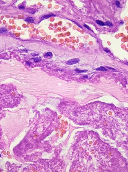
This Photo Shows The Pink Mucinous Matrix Of Atrial Myxoma, Myxoma Cells Arranged In Nests And Cords, And Local Bleeding.Magnify 1000x.
Image, 25.08MB, 4640 × 6226 jpg
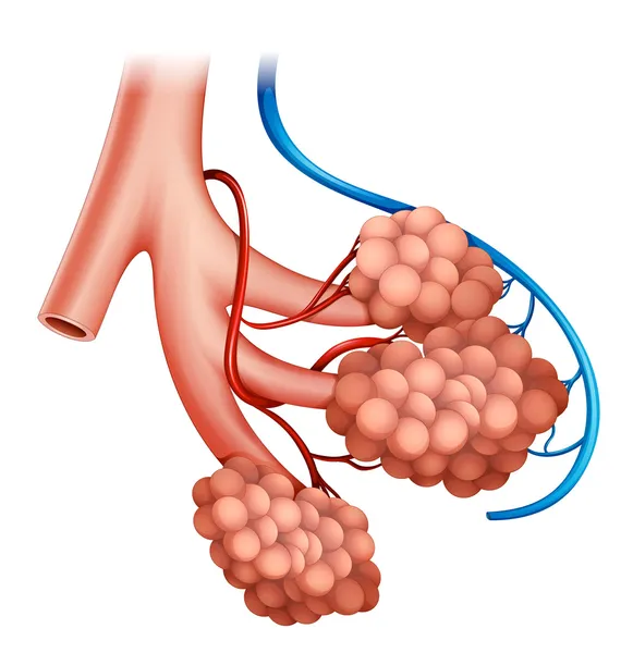

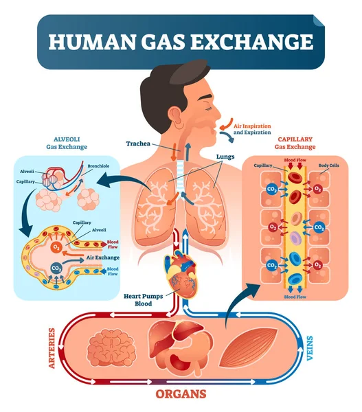
Human Gas Exchange System Vector Illustration. Oxygen Travel From Lungs To Heart, To All Body Cells And Back To Lungs As CO2.
Vector, 6.72MB, 5230 × 5951 eps

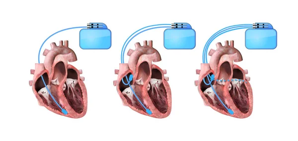
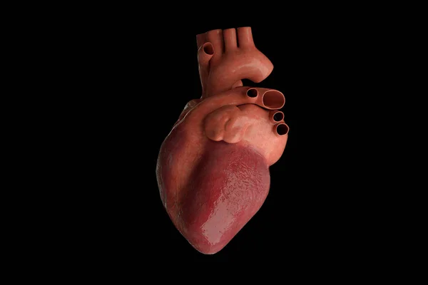
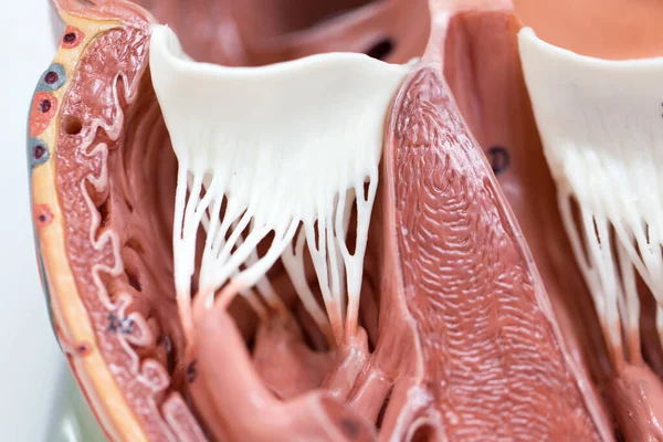
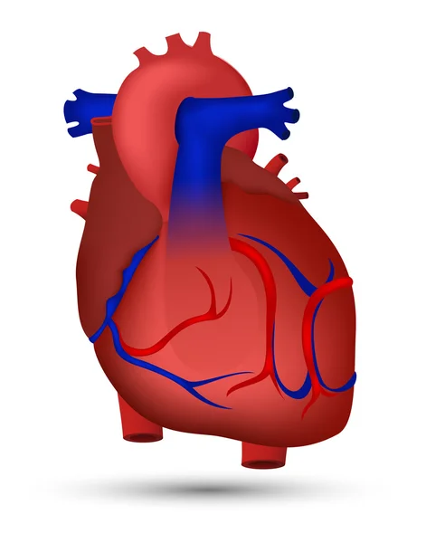
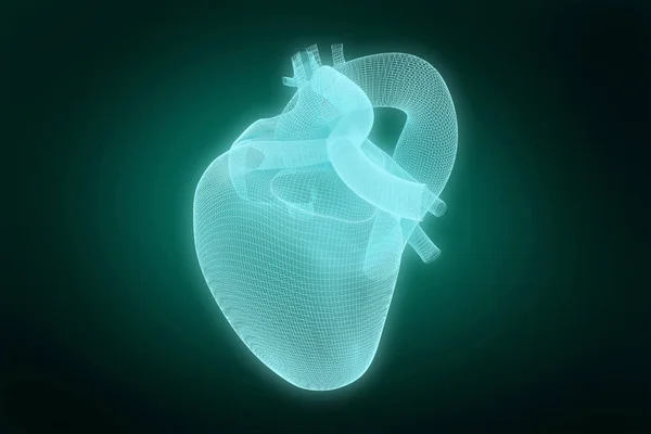
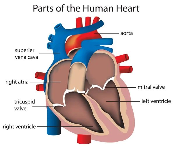
Heart And Cardiac Pacemaker On White Background 3d Render, Illustration Heart Anatomy, Section, Right And Left Ventricle, Atria, Valves
Image, 2.97MB, 5023 × 2392 jpg

Previous << Page 2 >> Next