Home
Images
Vectors
Videos
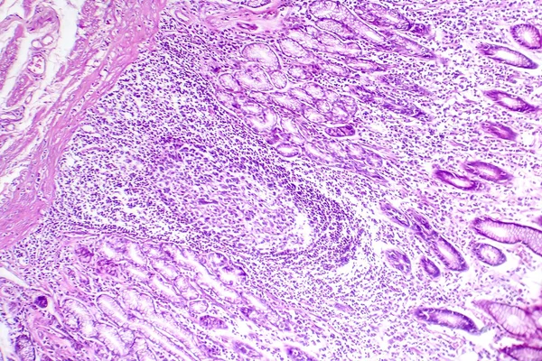
Stock image This photo shows the pink mucinous matrix of atrial myxoma, myxoma cells arranged in nests and cords, and local bleeding.Magnify 1000x.
Published:
Jul.20, 2024 16:26:39
Author:
asia11m
Views:
0
Downloads:
0
File type:
image
/
jpg
File size:
23.48 MB
Orginal size:
4640
x
6267
px
Available sizes:
S
(370 x 500)
M
(740 x 1000)
L
(1481 x 2000)
XL
(4640 x 6267)
EL0
(4640 x 6267)
Level:
beginner
Download Image
Image topics:
Photography
Human
Medicine
Illness
Heart
Cell
Disease
Cells
Congestion
Anatomy
Mucus
Tumor
Bleeding
Inflammation
Organs
Atrium
Infarction
Pathology
Tuberculosis
Serous
Nuclei
Biopsy
Glands
Tumors
Histology
Ischemia
Cytoplasm
Cross Section
Human Body
Necrosis
Stroma
Red Blood Cells
Histological
Micrography
Cardiac Surgery
Benign Tumors
Malignant Tumors
Cardiac Ultrasound
Fibrous Tissue
Dense Bodies
Hemosiderin
Myxoma
Left And Right Atria
Gandy Gamna Nodules
Myxoma Cells
Gandy Gamna Bodies
Calcium Deposition
Atrial Myxoma
Basic Medicine
Similar stock images
Gandy-Gamna Bodies Are Pathological Changes Involving Hemosiderosin And Calcium Salt Deposition Produced By Red Blood Cell Decomposition And Fibrous Tissue Encapsulation.
6500 × 7799
This Pathologic Photograph Highlights The Atrium Myxoma Cell Nest. Atrial Myxoma Is A Benign Tumor Of The Heart.
5500 × 7384
Photomicrograph Of Intestinal Metaplasia, Displaying Transformation Of Stomach Lining Cells Into Intestinal-like Cells.
6099 × 4067
Anatomy And Histological Ovary And Testis Human Cells Under Microscope.
6720 × 4480
Structure Of Tissue Of Spleen Human, Liver Human And Kidney Human Under The Microscope In Lab.
6720 × 4480
Chronic Nephritis, Light Micrograph, Photo Under Microscope
4252 × 2835
Breast Cancer, Light Micrograph, Photo Under Microscope
4438 × 2959
Ovarian Mucinous Cystadenoma, A Benign Tumor Of Ovary, Light Micrograph, Photo Under Microscope
4436 × 2957