
Stock image Cell Nuclei


Microscopic Image Of Onion Root Tip Cells Undergoing Mitosis. Anaphases And Metaphases. 1000x Magnified
Image, 2.36MB, 2592 × 1944 jpg

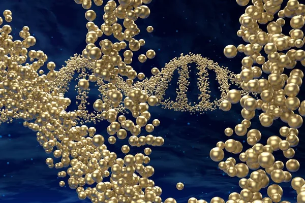
Gold Nucleotides In The DNA Molecule (human Genome) Double Helix Chain With Nebula Background - Through View 3d Illustration
Image, 15.89MB, 15000 × 10000 jpg


Microscopic Image Of Onion Root Tip Cells Undergoing Mitosis. Anaphases, Telophases And Metaphases. 1000x Magnified.
Image, 2.37MB, 2592 × 1944 jpg

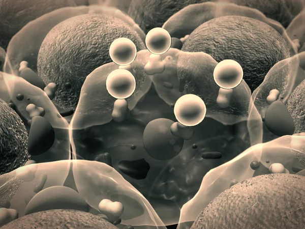
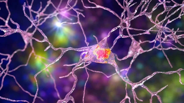
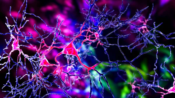
Neurons, Brain Cells Located In Amygdala, 3D Illustration. Amygdalas Are Clusters Of Nuclei Within The Temporal Lobes, Part Of The Limbic System, Their Neurons Play Role In Memory, Emotions
Image, 10.18MB, 7200 × 4050 jpg

Neurons, Brain Cells Located In Amygdala, 3D Illustration. Amygdalas Are Clusters Of Nuclei Within The Temporal Lobes, Part Of The Limbic System, Their Neurons Play Role In Memory, Emotions
Image, 8.86MB, 7200 × 4050 jpg

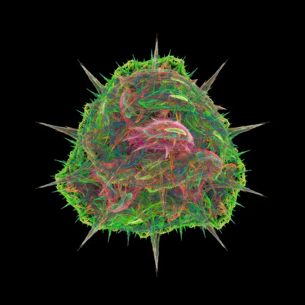
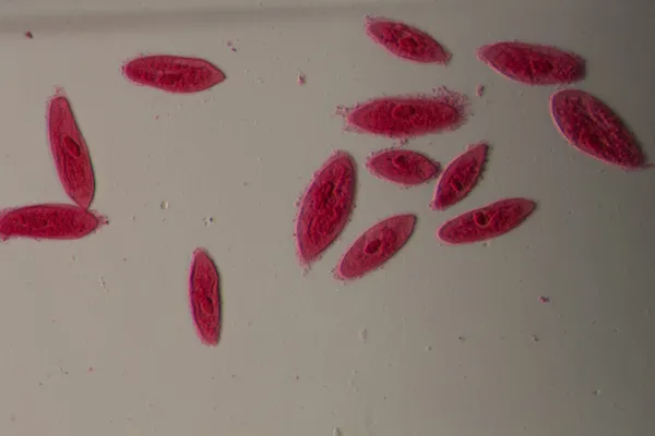
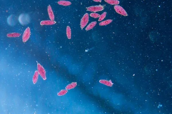
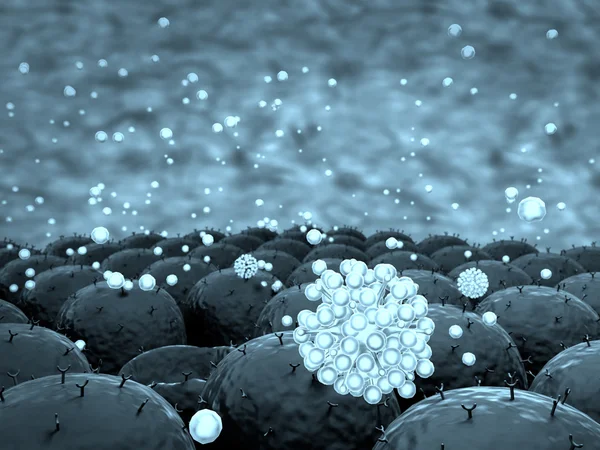
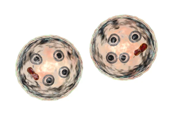
Cysts Of Entamoeba Coli Protozoan, 3D Illustration. E. Coli Is A Non-pathogenic Ameba, Its Cyst Is 15-25 Mkm, Has 8 Nuclei And Chromatoidal Bar Elongated With Splintered Ends (shown Red)
Image, 11.01MB, 5761 × 5761 jpg


Education Chart Of Biology For Germination Of Pollen On Stigma Diagram
Vector, 0.65MB, 5000 × 5000 eps
Amygdala In The Brain, And Closeup View Of Amygdala Neurons, 3D Illustration. Two Almond-shaped Clusters Of Nuclei Within Temporal Lobes, Part Of The Limbic System, Play Role In Memory And Emotions
Image, 11.08MB, 7111 × 4000 jpg
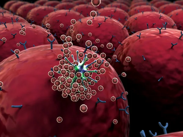
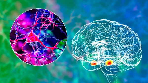
Caudate Nuclei In Human Brain And Its Neurons, 3D Illustration. The Caudate Nucleus Is A Component Of The Basal Ganglia, It Plays Role In Choreas, Neurodegenerative And Other Brain Diseases
Image, 16.77MB, 8157 × 5438 jpg

Caudate Nuclei In Human Brain And Its Neurons, 3D Illustration. The Caudate Nucleus Is A Component Of The Basal Ganglia, It Plays Role In Choreas, Neurodegenerative And Other Brain Diseases
Image, 12.8MB, 8157 × 5438 jpg
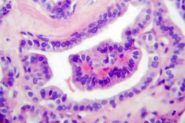
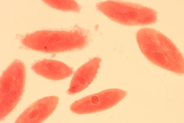
Papillary Thyroid Carcinoma, Light Micrograph, Photo Under Microscope. The Most Common Type Of Thyroid Cancer
Image, 3.3MB, 4126 × 2751 jpg

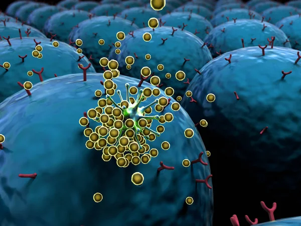
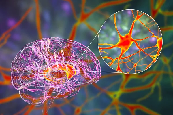
Destruction Of Neurons Of The Caudate Nucleus, Conceptual 3D Illustration. Caudate Nucleus Belongs To The Brain Basal Ganglia, Its Neurons Are Damaged In Huntingon's Disease And Other Choreas
Image, 16.49MB, 7996 × 5331 jpg
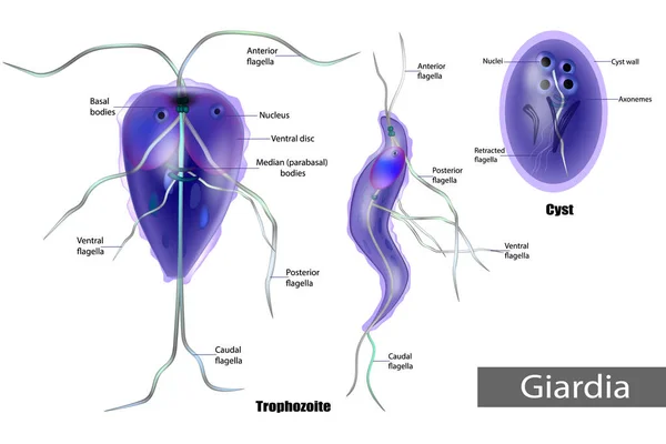
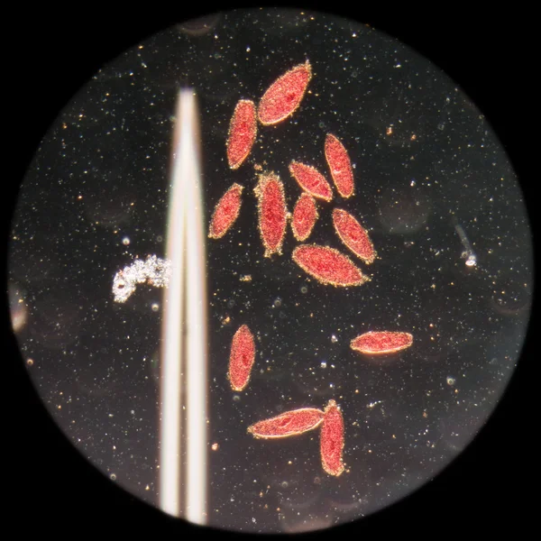
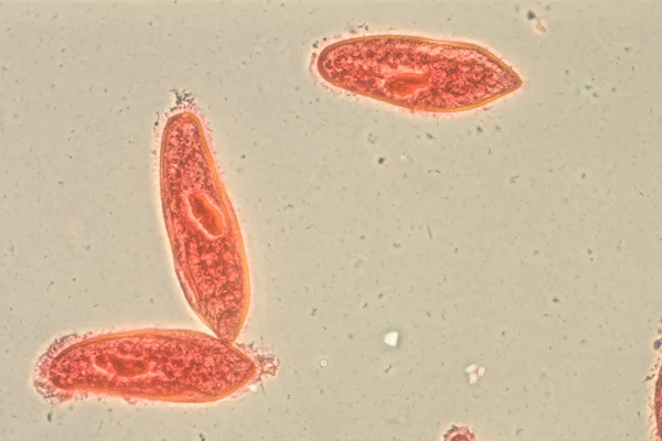
Giardia Anaerobic Flagellated Protozoan Parasites Of The Phylum Metamonada. The Structure Of Giardia Lamblia Of Cyst And Trophozoite. Giardiasis.
Vector, 10.88MB, 6000 × 4000 eps
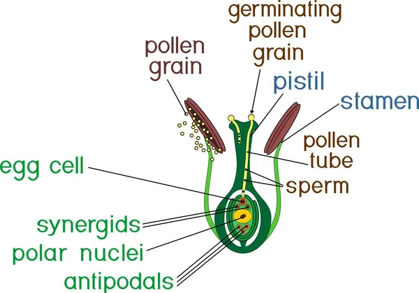
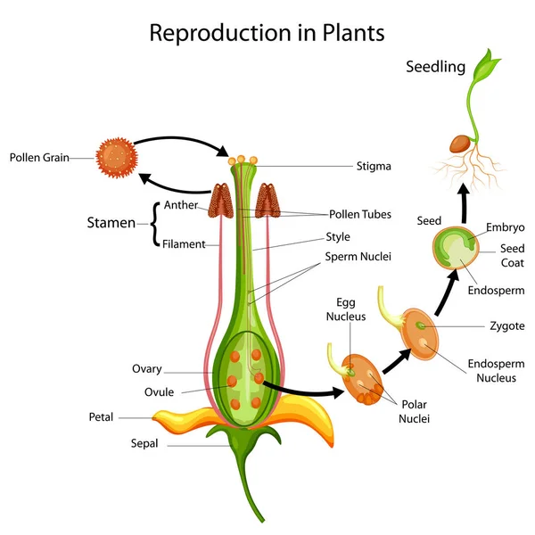
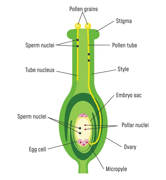
Structure Of Pistil And Stamens In The Section At The Time Of Double Fertilization
Vector, 0.25MB, 5000 × 3503 ai

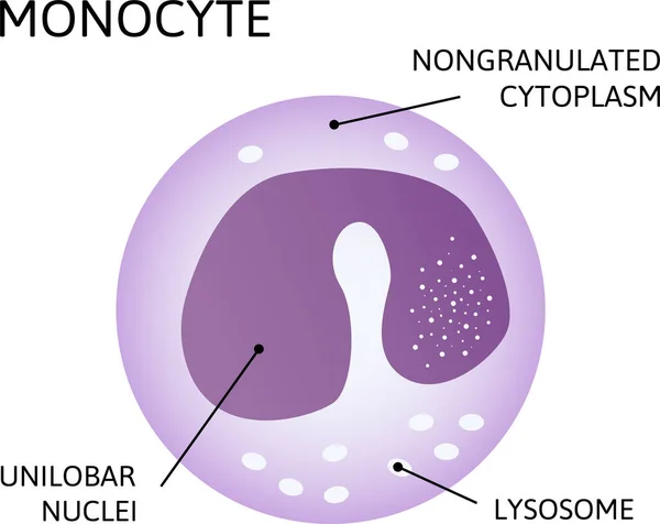
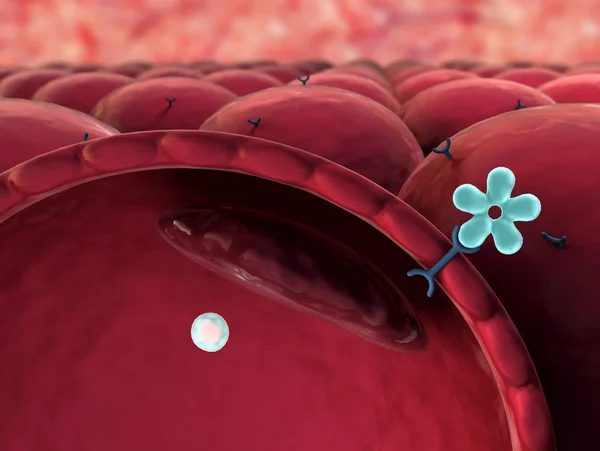
Monocytes, Type Of Leukocyte, White Blood Cell. Consist Of Nongranulated Cytoplasm, Unilobar Nuclei And Lysosome. Vector Medical Illustration
Vector, 0.39MB, 5407 × 4300 eps
Neurons, Brain Cells Located In Amygdala, 3D Illustration. Amygdalas Are Clusters Of Nuclei Within The Temporal Lobes, Part Of The Limbic System, Their Neurons Play Role In Memory, Emotions
Image, 10.04MB, 7200 × 4050 jpg
Cysts Of Entamoeba Histolityca, 3D Illustration. Parasitic Ameba, The Causative Agent Of Amebic Dysentery And Extraintestinal Amebiasis
Image, 3.99MB, 6000 × 4000 jpg

Neurons, Brain Cells Located In Amygdala, 3D Illustration. Amygdalas Are Clusters Of Nuclei Within The Temporal Lobes, Part Of The Limbic System, Their Neurons Play Role In Memory, Emotions
Image, 8.06MB, 7200 × 4050 jpg

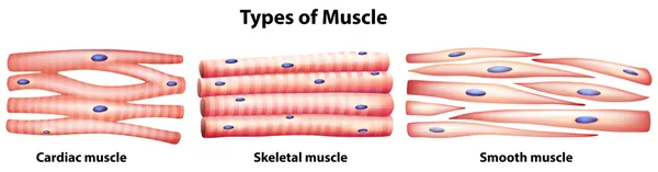
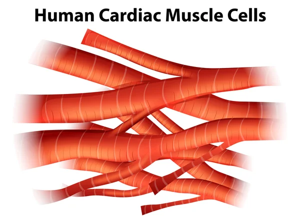
Muscle Membrane Or Sarcolemma Anatomical Layers Structure Outline Diagram
Vector, 7.08MB, 5000 × 3913 eps


Gold Nucleotides In The DNA Molecule (human Genome) Double Helix Chain With Nebula Background - Isometric View 3d Illustration
Image, 12.93MB, 15000 × 10000 jpg
Human Chromosomes (23 + X, Y) Structures Made Of Protein And A Single Molecule Of DNA - Closeup View 3d Illustration
Image, 10.04MB, 10000 × 6600 jpg

Page 1 >> Next