photo free
Home Images Vectors Videos

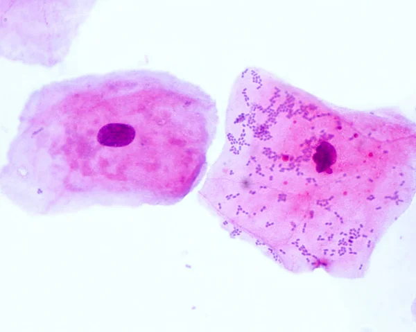
Stock image Histology

stock image African female tech, scientists white coat and gloves do pcr test, work with microscope in modern laboratory or research facility. Analysis of lung tissue from COVID-19 patients, clinical cases.

African Female Tech, Scientists White Coat And Gloves Do Pcr Test, Work With Microscope In Modern Laboratory Or Research Facility. Analysis Of Lung Tissue From COVID-19 Patients, Clinical Cases.

Image, 7.16MB, 3995 × 2804 jpg
stock image Adipose tissue

Adipose Tissue

Image, 10.4MB, 3840 × 3072 jpg
stock image biological cell isolated on whithe background microscope

Biological Cell Isolated On Whithe Background Microscope

Image, 5.88MB, 6000 × 4000 jpg
stock image Kidney. Collecting duct

Kidney. Collecting Duct

Image, 12.93MB, 3840 × 3072 jpg
stock image Mast cell

Mast Cell

Image, 17.59MB, 3840 × 3072 jpg
stock image Cancer cell in the moment that divides, 3d illustration

Cancer Cell In The Moment That Divides, 3d Illustration

Image, 6.82MB, 5001 × 3865 jpg
stock image Coccidiosis, coccidia in liver, light micrograph, photo under microscope

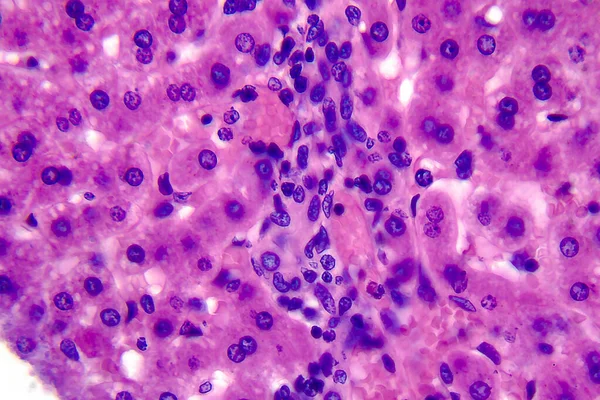
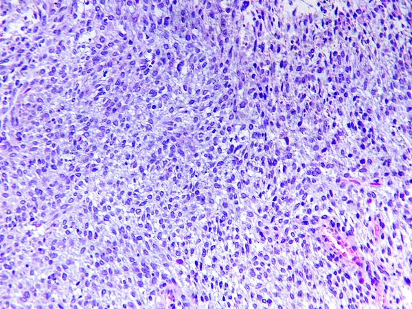
Coccidiosis, Coccidia In Liver, Light Micrograph, Photo Under Microscope

Image, 4.14MB, 4055 × 2703 jpg
stock image Endometrial adenocarcinoma, light micrograph, photo under microscope

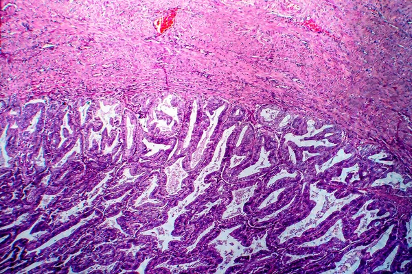
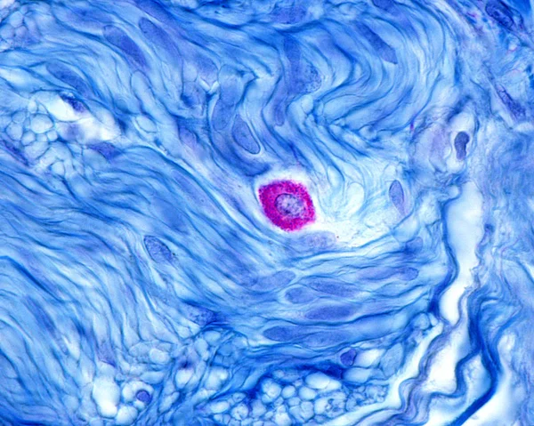
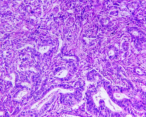
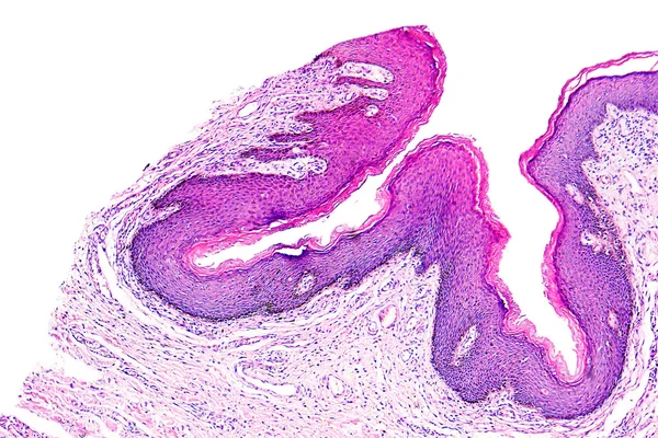
Endometrial Adenocarcinoma, Light Micrograph, Photo Under Microscope

Image, 15.15MB, 4060 × 2707 jpg
stock image The words CANCER DIAGNOSIS written on a white notepad on a blue background near a stethoscope, syringe, electronic thermometer and pills. Medical concept

The Words CANCER DIAGNOSIS Written On A White Notepad On A Blue Background Near A Stethoscope, Syringe, Electronic Thermometer And Pills. Medical Concept

Image, 5.23MB, 5287 × 3500 jpg
stock image Squamous cell carcinoma of a human

Squamous Cell Carcinoma Of A Human

Image, 4.72MB, 2240 × 1792 jpg
stock image Scientist is preparing a tumor slide. Microscopy of cytopathology slides and pathology.

Scientist Is Preparing A Tumor Slide. Microscopy Of Cytopathology Slides And Pathology.

Image, 12.02MB, 6000 × 4000 jpg
stock image Tissue sections, battery of jars with fixed tissue samples on gl

Tissue Sections, Battery Of Jars With Fixed Tissue Samples On Gl

Image, 1.86MB, 4824 × 3216 jpg
stock image Chronic nephritis, light micrograph, photo under microscope. High magnification

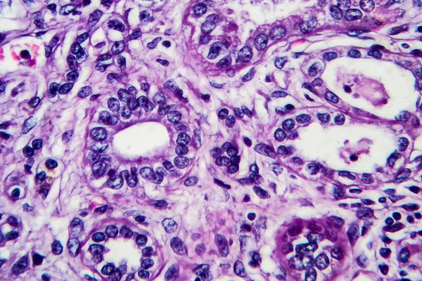
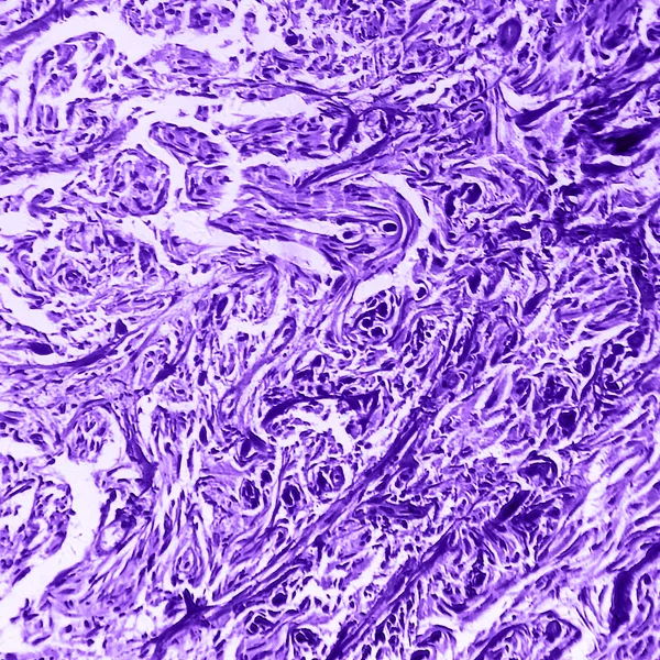
Chronic Nephritis, Light Micrograph, Photo Under Microscope. High Magnification

Image, 9.38MB, 4546 × 3031 jpg
stock image Gastric adenocarcinoma, light micrograph

Gastric Adenocarcinoma, Light Micrograph

Image, 9.22MB, 4216 × 2811 jpg
stock image Pancreas section organic background

Pancreas Section Organic Background

Image, 16.6MB, 5032 × 3358 jpg
stock vector Different types of epithelial tissue collection.

Different Types Of Epithelial Tissue Collection.

Vector, 2.39MB, 5000 × 5671 eps
stock image Squamous epithelial cells

Squamous Epithelial Cells

Image, 6.41MB, 3840 × 3072 jpg
stock image Taste buds

Taste Buds

Image, 11.36MB, 3840 × 3072 jpg
stock vector Epithelial cells vector illustration. Medical location and meaning diagram.

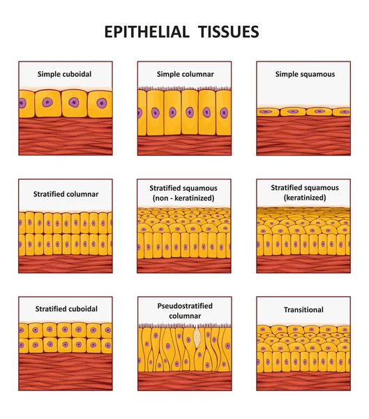
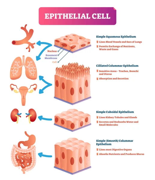
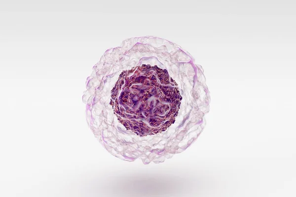
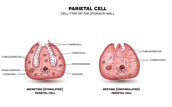
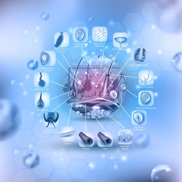
Epithelial Cells Vector Illustration. Medical Location And Meaning Diagram.

Vector, 8.27MB, 3750 × 4354 eps
stock image Heart muscle, light micrograph

Heart Muscle, Light Micrograph

Image, 4.46MB, 1918 × 4384 jpg
stock image Male scientist looking at slides under the microscope

Male Scientist Looking At Slides Under The Microscope

Image, 8.38MB, 3497 × 5245 jpg
stock image Scientists in laboratory regarding sample in petri dish

Scientists In Laboratory Regarding Sample In Petri Dish

Image, 5.38MB, 5760 × 3840 jpg
stock image Group of food laboratory researchers comparing bacteria cultures

Group Of Food Laboratory Researchers Comparing Bacteria Cultures

Image, 5.04MB, 5760 × 3840 jpg
stock image Brain astrocytoma of a human

Brain Astrocytoma Of A Human

Image, 5.55MB, 2004 × 2004 jpg
stock image Fibrosarcoma of a human

Fibrosarcoma Of A Human

Image, 5.17MB, 2312 × 1734 jpg
stock image Gastric adenocarcinoma of a human

Gastric Adenocarcinoma Of A Human

Image, 6.31MB, 2240 × 1792 jpg
stock vector Parietal cell of stomach wall, located in the gastric glands sec

Parietal Cell Of Stomach Wall, Located In The Gastric Glands Sec

Vector, 14.74MB, 6100 × 3810 eps
stock vector Cell division background

Cell Division Background

Vector, 11.22MB, 3252 × 2215 eps
stock vector Skin anatomy round shape, detailed illustration

Skin Anatomy Round Shape, Detailed Illustration

Vector, 11.15MB, 5000 × 5000 eps
stock image Light micrograph of a kidney

Light Micrograph Of A Kidney

Image, 1.69MB, 3024 × 2016 jpg
stock image Ovarian follicle

Ovarian Follicle

Image, 2.7MB, 3930 × 2620 jpg
stock image Scientist works with the microscope slide in lab

Scientist Works With The Microscope Slide In Lab

Image, 7.7MB, 5184 × 3456 jpg
stock image Skin papilloma of a human

Skin Papilloma Of A Human

Image, 6.26MB, 2775 × 1850 jpg
stock image micrograph plant root tip tissue cell

Micrograph Plant Root Tip Tissue Cell

Image, 7.16MB, 3107 × 3107 jpg
stock image Lab technician examines scrapings on glass slides

Lab Technician Examines Scrapings On Glass Slides

Image, 13.93MB, 5760 × 3840 jpg
stock image Male scientist or tech with dark hair and brown eyes works in sc

Male Scientist Or Tech With Dark Hair And Brown Eyes Works In Sc

Image, 8.95MB, 6645 × 4435 jpg
stock vector carcinoid cancer medical zebra ribbon treatment health disease

Carcinoid Cancer Medical Zebra Ribbon Treatment Health Disease

Vector, 0.29MB, 5000 × 5000 eps
stock vector Microscope icon. Medical laboratory equipment.

Microscope Icon. Medical Laboratory Equipment.

Vector, 2.05MB, 5556 × 5556 eps
stock vector Microscope icon. Medical laboratory equipment.

Microscope Icon. Medical Laboratory Equipment.

Vector, 0.96MB, 5556 × 5556 eps
stock image Health care professionals.

Health Care Professionals.

Image, 2.42MB, 3000 × 2000 jpg
stock image Multicellular Epithelium structure

Multicellular Epithelium Structure

Image, 4.64MB, 4096 × 4096 jpg
stock image Notochord formation illustration

Notochord Formation Illustration

Image, 2.39MB, 3840 × 2160 jpg
stock image Notochord formation illustration

Notochord Formation Illustration

Image, 2.97MB, 3840 × 2160 jpg
stock image Human lung affected by an acute bronchopneumonia, commonly a hospital-acquired bacterial pneumonia. The lumen of alveoli is occupied by liquid of oedema which contains acute inflammatory infiltrates (with predominance of neutrophil granulocytes). On

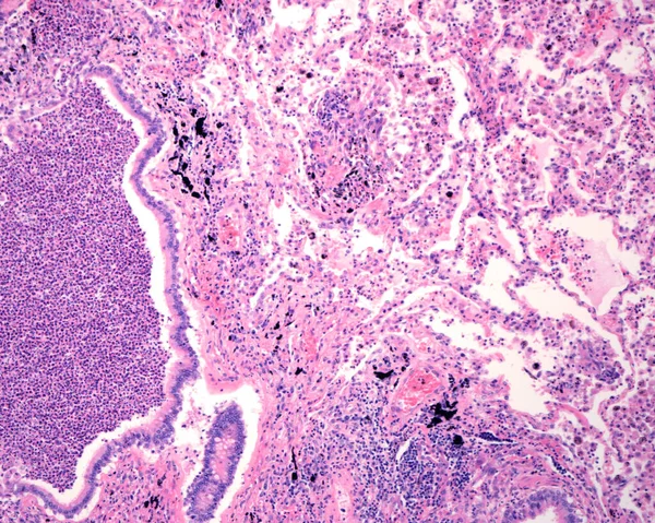
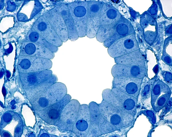
Human Lung Affected By An Acute Bronchopneumonia, Commonly A Hospital-acquired Bacterial Pneumonia. The Lumen Of Alveoli Is Occupied By Liquid Of Oedema Which Contains Acute Inflammatory Infiltrates (with Predominance Of Neutrophil Granulocytes). On

Image, 11.33MB, 3840 × 3072 jpg
stock vector Microscope icon. Medical laboratory equipment.

Microscope Icon. Medical Laboratory Equipment.

Vector, 0.89MB, 5556 × 5556 eps
stock vector Liver disease, variety of illnesses can affect the liver

Liver Disease, Variety Of Illnesses Can Affect The Liver

Vector, 12.03MB, 4600 × 3396 eps
stock image Science Researcher

Science Researcher

Image, 3.4MB, 7575 × 4000 jpg
stock image Under high magnification microscopic view of vagina

Under High Magnification Microscopic View Of Vagina

Image, 7.77MB, 5700 × 3773 jpg
stock image Developing long bone. Growth plate

Developing Long Bone. Growth Plate

Image, 12.4MB, 3421 × 2800 jpg
stock image Tools for histochemical analysis of patient tissue

Tools For Histochemical Analysis Of Patient Tissue

Image, 2.04MB, 3462 × 2300 jpg
Page 1 >> Next