photo free
Home Images Vectors Videos

Stock image Histology page 2

stock vector Parietal cell of stomach wall, located in the gastric glands sec

Parietal Cell Of Stomach Wall, Located In The Gastric Glands Sec

Vector, 14.74MB, 6100 × 3810 eps
stock vector Cell division background

Cell Division Background

Vector, 11.22MB, 3252 × 2215 eps
stock vector Skin anatomy round shape, detailed illustration

Skin Anatomy Round Shape, Detailed Illustration

Vector, 11.15MB, 5000 × 5000 eps
stock image Light micrograph of a kidney

Light Micrograph Of A Kidney

Image, 1.69MB, 3024 × 2016 jpg
stock image Ovarian follicle

Ovarian Follicle

Image, 2.7MB, 3930 × 2620 jpg
stock image micrograph plant root tip tissue cell

Micrograph Plant Root Tip Tissue Cell

Image, 7.16MB, 3107 × 3107 jpg
stock image Lab technician examines scrapings on glass slides

Lab Technician Examines Scrapings On Glass Slides

Image, 13.93MB, 5760 × 3840 jpg
stock image Male scientist or tech with dark hair and brown eyes works in sc

Male Scientist Or Tech With Dark Hair And Brown Eyes Works In Sc

Image, 8.95MB, 6645 × 4435 jpg
stock vector carcinoid cancer medical zebra ribbon treatment health disease

Carcinoid Cancer Medical Zebra Ribbon Treatment Health Disease

Vector, 0.29MB, 5000 × 5000 eps
stock vector Microscope icon. Medical laboratory equipment.

Microscope Icon. Medical Laboratory Equipment.

Vector, 2.05MB, 5556 × 5556 eps
stock vector Microscope icon. Medical laboratory equipment.

Microscope Icon. Medical Laboratory Equipment.

Vector, 0.96MB, 5556 × 5556 eps
stock image Health care professionals.

Health Care Professionals.

Image, 2.42MB, 3000 × 2000 jpg
stock image Multicellular Epithelium structure

Multicellular Epithelium Structure

Image, 4.64MB, 4096 × 4096 jpg
stock image Notochord formation illustration

Notochord Formation Illustration

Image, 2.39MB, 3840 × 2160 jpg
stock image Notochord formation illustration

Notochord Formation Illustration

Image, 2.97MB, 3840 × 2160 jpg
stock image Human lung affected by an acute bronchopneumonia, commonly a hospital-acquired bacterial pneumonia. The lumen of alveoli is occupied by liquid of oedema which contains acute inflammatory infiltrates (with predominance of neutrophil granulocytes). On

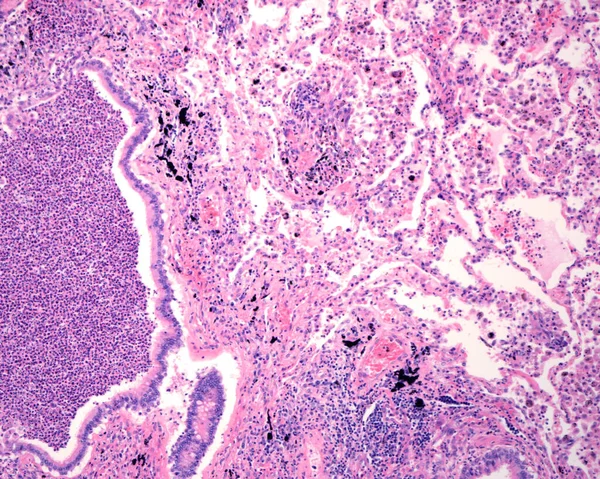
Human Lung Affected By An Acute Bronchopneumonia, Commonly A Hospital-acquired Bacterial Pneumonia. The Lumen Of Alveoli Is Occupied By Liquid Of Oedema Which Contains Acute Inflammatory Infiltrates (with Predominance Of Neutrophil Granulocytes). On

Image, 11.33MB, 3840 × 3072 jpg
stock vector Microscope icon. Medical laboratory equipment.

Microscope Icon. Medical Laboratory Equipment.

Vector, 0.89MB, 5556 × 5556 eps
stock vector Liver disease, variety of illnesses can affect the liver

Liver Disease, Variety Of Illnesses Can Affect The Liver

Vector, 12.03MB, 4600 × 3396 eps
stock image Science Researcher

Science Researcher

Image, 3.4MB, 7575 × 4000 jpg
stock image Developing long bone. Growth plate

Developing Long Bone. Growth Plate

Image, 12.4MB, 3421 × 2800 jpg
stock image Tools for histochemical analysis of patient tissue

Tools For Histochemical Analysis Of Patient Tissue

Image, 2.04MB, 3462 × 2300 jpg
stock image science medical anthropotomy physiology microscopic human small

Science Medical Anthropotomy Physiology Microscopic Human Small

Image, 4.49MB, 2635 × 2635 jpg
stock image microscopic section of human kidney tissue

Microscopic Section Of Human Kidney Tissue

Image, 7.29MB, 2839 × 2839 jpg
stock image Array of blood samples for microscopy and biopsy tissue on blue

Array Of Blood Samples For Microscopy And Biopsy Tissue On Blue

Image, 5.04MB, 4116 × 2744 jpg
stock image Training model plant stem isolated on white background

Training Model Plant Stem Isolated On White Background

Image, 4.06MB, 4134 × 3000 jpg
stock image Endoscopic medical device in the hands of a surgeon's. Endoscopist is ready to work with the patient.

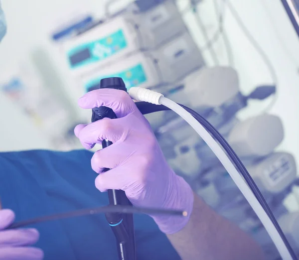
Endoscopic Medical Device In The Hands Of A Surgeon's. Endoscopist Is Ready To Work With The Patient.

Image, 1.69MB, 2869 × 2500 jpg
stock image PC-3 human prostate cancer cells stained with blue Coomassie, un

PC-3 Human Prostate Cancer Cells Stained With Blue Coomassie, Un

Image, 6.34MB, 4184 × 2789 jpg
stock image Stop Preeclampsia on Open Hand.

Stop Preeclampsia On Open Hand.

Image, 2.53MB, 2922 × 3092 jpg
stock vector Egg of the liver fluke. infographics. Vector illustration on isolated background.

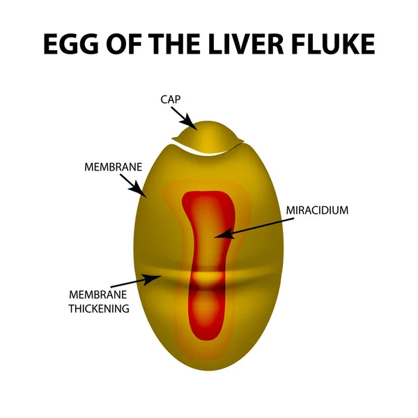
Egg Of The Liver Fluke. Infographics. Vector Illustration On Isolated Background.

Vector, 1.68MB, 5000 × 5000 eps
stock image Close up human cells with microscope.

Close Up Human Cells With Microscope.

Image, 2.35MB, 3200 × 2304 jpg
stock image The study Plant tissue of under the microscope for classroom education.

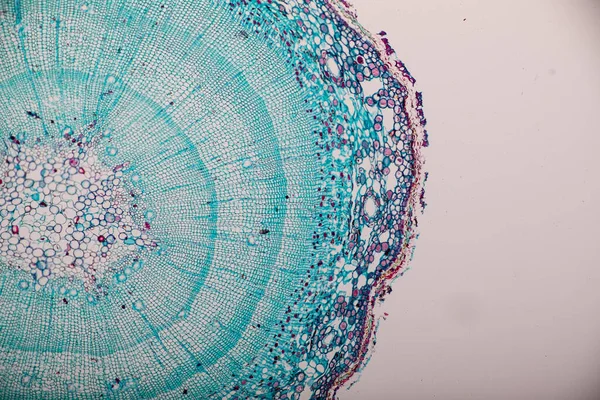
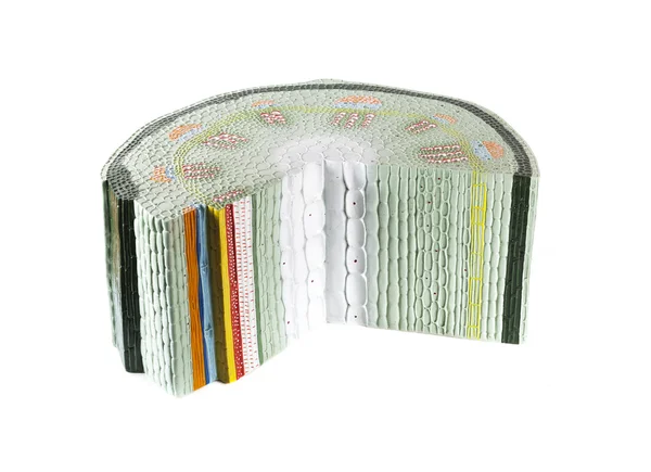
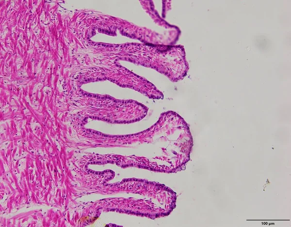
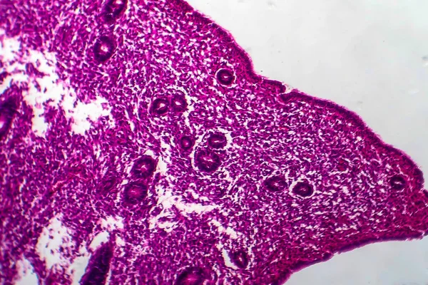
The Study Plant Tissue Of Under The Microscope For Classroom Education.

Image, 20.8MB, 6720 × 4480 jpg
stock image Animal tissue samples under the microscope in Lab.

Animal Tissue Samples Under The Microscope In Lab.

Image, 0.85MB, 2448 × 1920 jpg
stock image Cell Division and Cell Cycle under the microscope.

Cell Division And Cell Cycle Under The Microscope.

Image, 16.67MB, 6720 × 4480 jpg
stock image Root tip of Onion and Mitosis cell in the Root tip of Onion under a microscope.

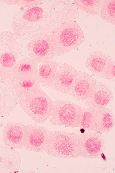
Root Tip Of Onion And Mitosis Cell In The Root Tip Of Onion Under A Microscope.

Image, 4.92MB, 2560 × 3840 jpg
stock image  Concept of stomach puncture or gastrointestinal perforation. Hand of surgeon pierces wall of model of human stomach for therapeutic purposes or for biopsy tissue analysis by histology or cytology

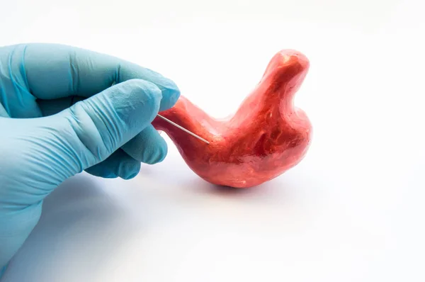
Concept Of Stomach Puncture Or Gastrointestinal Perforation. Hand Of Surgeon Pierces Wall Of Model Of Human Stomach For Therapeutic Purposes Or For Biopsy Tissue Analysis By Histology Or Cytology

Image, 5.08MB, 6016 × 4000 jpg
stock image Elastic cartilage of human outer ear

Elastic Cartilage Of Human Outer Ear

Image, 3.09MB, 3849 × 2566 jpg
stock image Intestinal adenomas, light micrograph

Intestinal Adenomas, Light Micrograph

Image, 2.82MB, 4042 × 2695 jpg
stock image Chronic superficial gastritis, light micrograph

Chronic Superficial Gastritis, Light Micrograph

Image, 5.62MB, 3701 × 2467 jpg
stock image Caseous pneumonia, light micrograph

Caseous Pneumonia, Light Micrograph

Image, 3.37MB, 4165 × 2777 jpg
stock image Uterus adenofibroma, light micrograph, photo under microscope. A rare benign tumor of the uterus composed of glandular and fibrous tissues

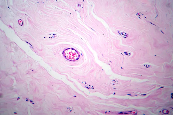
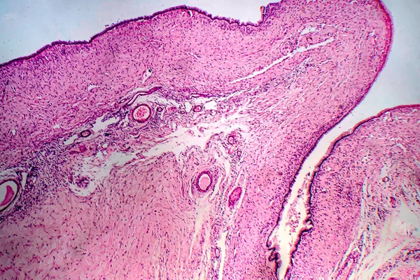
Uterus Adenofibroma, Light Micrograph, Photo Under Microscope. A Rare Benign Tumor Of The Uterus Composed Of Glandular And Fibrous Tissues

Image, 9.96MB, 4602 × 3068 jpg
stock image Breast ductal carcinoma, light micrograph, photo under microscope

Breast Ductal Carcinoma, Light Micrograph, Photo Under Microscope

Image, 14.07MB, 4103 × 2735 jpg
stock image Chronic pyelonephritis, light micrograph, photo under microscope. High magnification

Chronic Pyelonephritis, Light Micrograph, Photo Under Microscope. High Magnification

Image, 10.19MB, 4832 × 3221 jpg
stock image Histopathology of diffuse sclerosing glomerulonephritis, light micrograph, photo under microscope

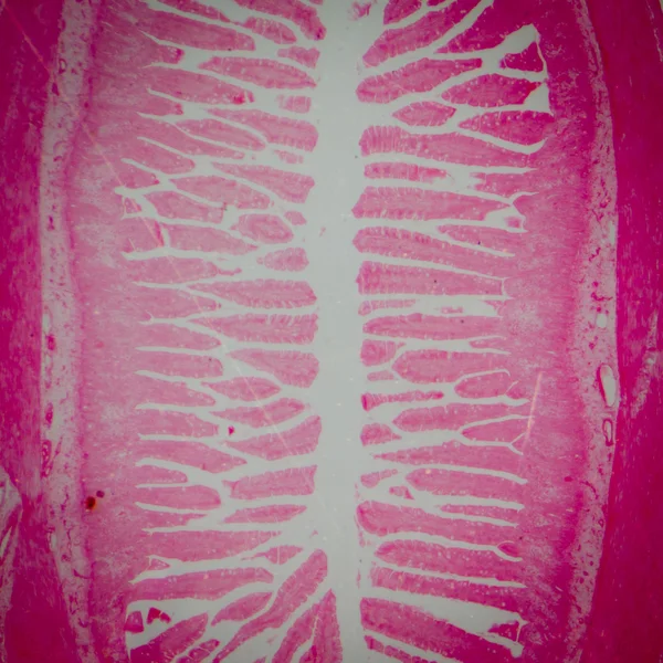
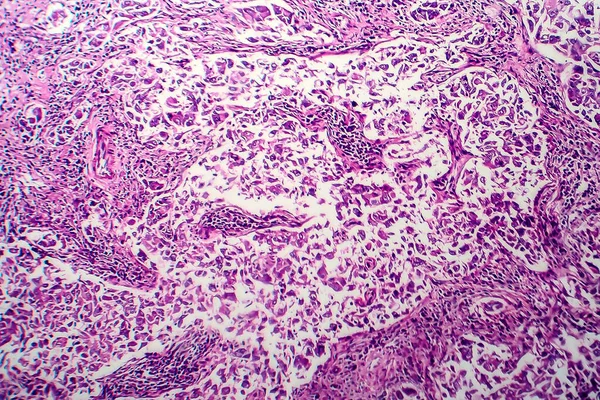
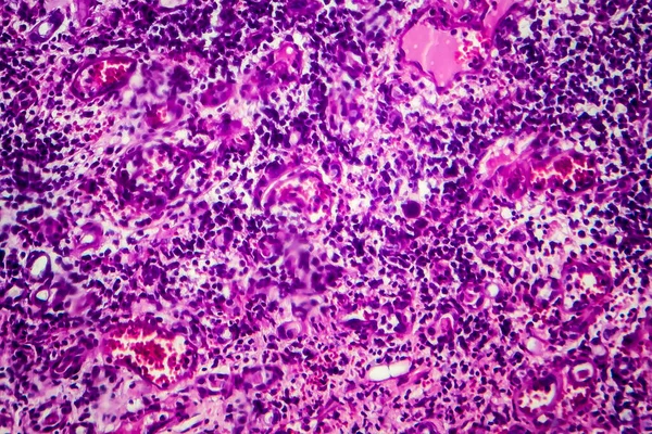
Histopathology Of Diffuse Sclerosing Glomerulonephritis, Light Micrograph, Photo Under Microscope

Image, 8.72MB, 4342 × 2895 jpg
stock image Tubular atrophy, light micrograph, photo under microscope. High magnification

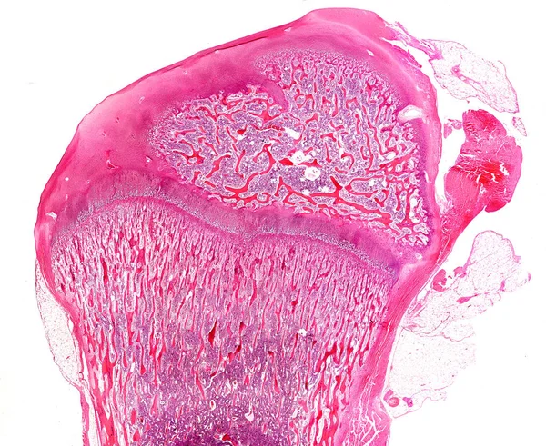
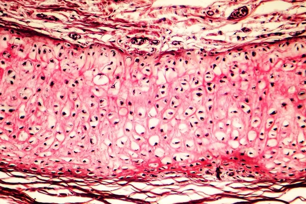
Tubular Atrophy, Light Micrograph, Photo Under Microscope. High Magnification

Image, 7.83MB, 3628 × 2419 jpg
stock image Stomach adenocarcinoma, gastric cancer, illustration and light micrograph

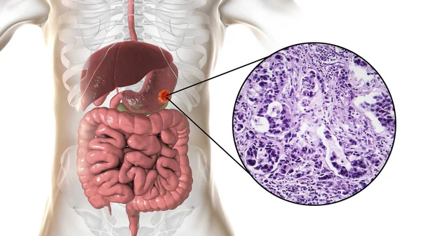
Stomach Adenocarcinoma, Gastric Cancer, Illustration And Light Micrograph

Image, 5.8MB, 5333 × 3000 jpg
stock image Ovarian cyst, light micrograph, photo under microscope

Ovarian Cyst, Light Micrograph, Photo Under Microscope

Image, 11.62MB, 3970 × 2647 jpg
stock image Endometrial hyperplasia, light micrograph, photo under microscope

Endometrial Hyperplasia, Light Micrograph, Photo Under Microscope

Image, 11.87MB, 4272 × 2848 jpg
stock image Histopathology of interstitial nephritis, light micrograph, photo under microscope

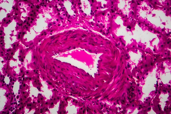
Histopathology Of Interstitial Nephritis, Light Micrograph, Photo Under Microscope

Image, 10.65MB, 3956 × 2637 jpg
stock image Human intestine with intestinal bacteria, 3D illustration

Human Intestine With Intestinal Bacteria, 3D Illustration

Image, 1.83MB, 4500 × 3000 jpg
stock image Liver with cirrhosis inside human body. 3D illustration and light micrograph of biliary cirrhosis

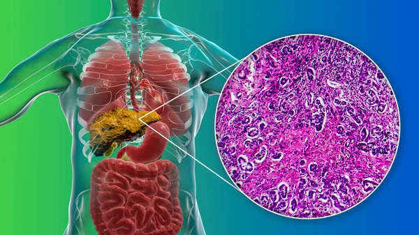
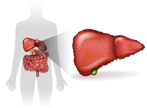
Liver With Cirrhosis Inside Human Body. 3D Illustration And Light Micrograph Of Biliary Cirrhosis

Image, 10.35MB, 5669 × 3189 jpg
Previous << Page 2 >> Next