
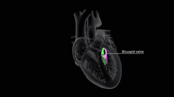
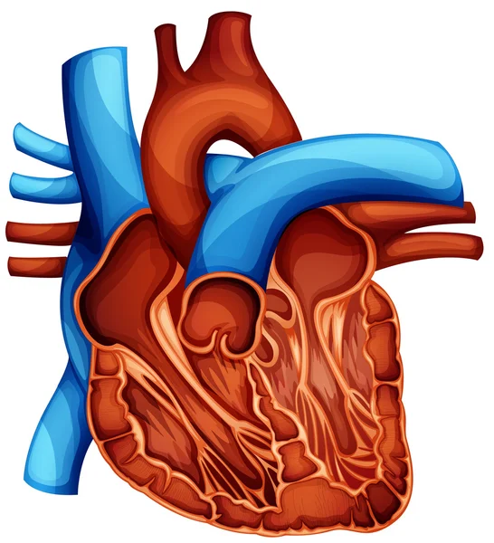
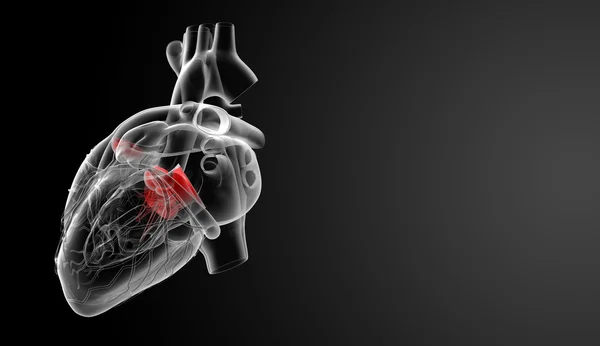
Stock image Bicuspid Valve

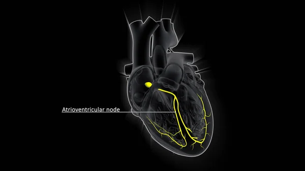
The AV Node, Which Controls The Heart Rate, Is One Of The Major Elements In The Cardiac Conduction System. The AV Node Serves As An Electrical Relay Station, Slowing The Electrical Current Sent By The Sinoatrial (SA) Node.
Image, 2.16MB, 3840 × 2160 jpg
The AV Node, Which Controls The Heart Rate, Is One Of The Major Elements In The Cardiac Conduction System. The AV Node Serves As An Electrical Relay Station, Slowing The Electrical Current Sent By The Sinoatrial (SA) Node.
Image, 1.15MB, 3840 × 2160 jpg
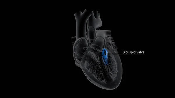
A Bicuspid Aortic Valve Is An Aortic Valve That Has Two Flaps (cusps) Instead Of Three. It May Cause A Narrowed Or Obstructed Aortic Valve Opening , Making It Difficult For The Heart To Pump Blood Into The Body's Main Artery (aorta).
Image, 0.95MB, 3840 × 2160 jpg
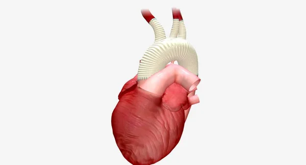
A Synthetic Graft Is An Artificial Tube That Allows The Oxygenated Blood To Flowfrom The Heart To The Rest Of The Body. 3D Rendering
Image, 3.17MB, 7258 × 3920 jpg
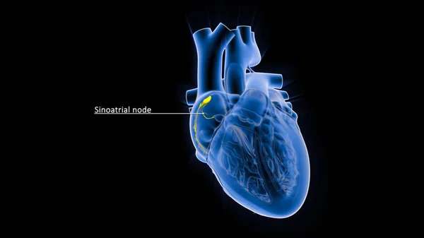
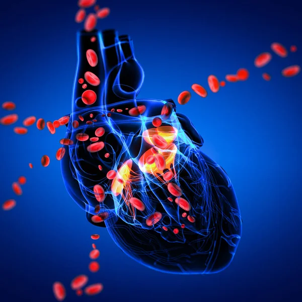
The Sinoatrial Node (also Known As The Sinuatrial Node, SA Node Or Sinus Node) Is A Group Of Cells Located In The Wall Of The Right Atrium Of The Heart.
Image, 1.32MB, 3840 × 2160 jpg
Hypertriglyceridemia Is A Common Condition Characterized By High Levels Of Triglycerides In The Bloodstream. 3D Render
Image, 5.46MB, 7258 × 3920 jpg
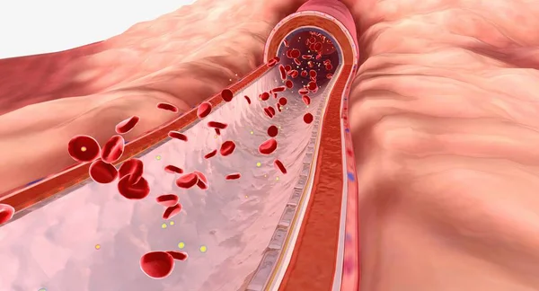
Lipids Are Types Of Fat That Travel Through The Bloodstream. 3D Render
Image, 15.08MB, 7258 × 3920 jpg
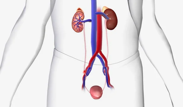
Angiotensin Receptor Neprilysin Inhibitors (ARNIs) Are A Combination Medication That Work By Helping The Kidney Move Extra Sodium Into The Ureter, Where It Is Flushed Away With Urine. 3D Rendering
Image, 14.49MB, 7258 × 3920 jpg

The AV Node, Which Controls The Heart Rate, Is One Of The Major Elements In The Cardiac Conduction System. The AV Node Serves As An Electrical Relay Station, Slowing The Electrical Current Sent By The Sinoatrial (SA) Node
Image, 1.59MB, 3840 × 2160 jpg
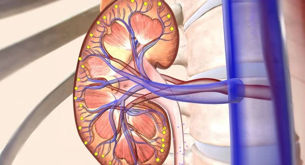
Kidney Stones Can Become Painful When Traveling Through The Urinary Tract But Do Not Usually Cause Lasting Damage. 3D Rendering
Image, 4.94MB, 7191 × 4200 jpg

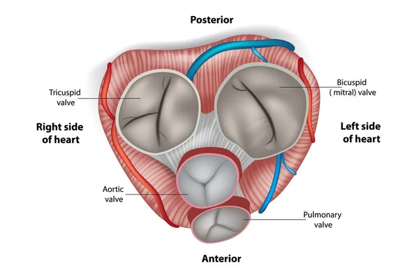
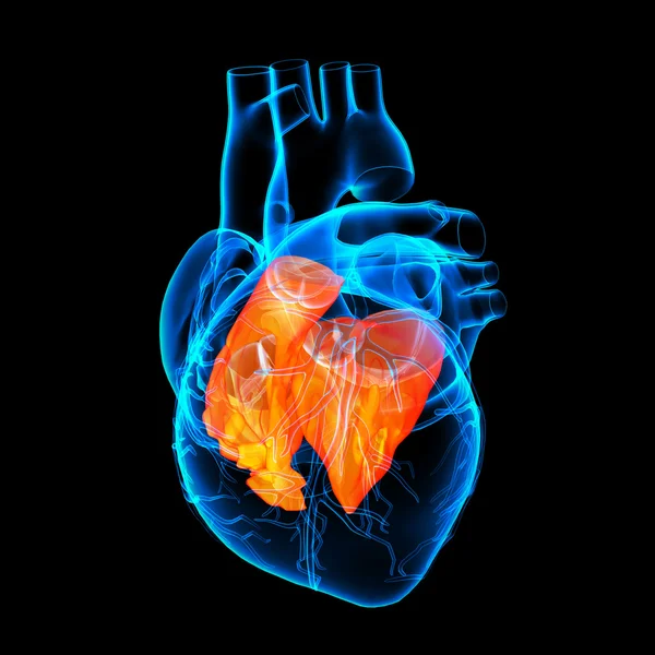
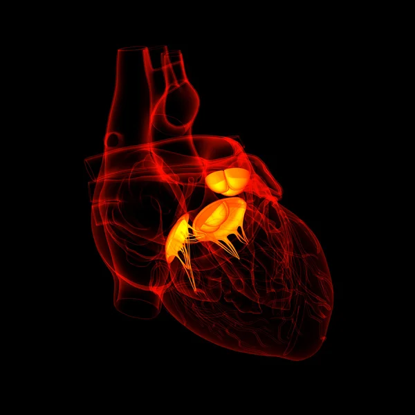
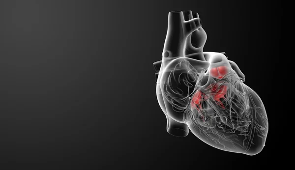
3d Illustration Human Heart Tricuspid And Bicuspid Valve For Medical Concept
Image, 5.53MB, 6000 × 3375 jpg
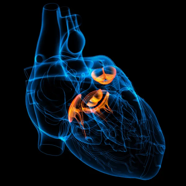
A Bicuspid Aortic Valve Is An Aortic Valve That Has Two Flaps (cusps) Instead Of Three. It May Cause A Narrowed Or Obstructed Aortic Valve Opening , Making It Difficult For The Heart To Pump Blood Into The Body's Main Artery (aorta).
Image, 0.96MB, 3840 × 2160 jpg

The SA (sinoatrial) Node Generates An Electrical Signal That Causes The Upper Heart Chambers To Contract. The Signal Then Passes Through The AV (atrioventricular) Node To The Lower Heart Chambers (ventricles), Causing Them To Contract, Or Pump.
Image, 1.19MB, 3840 × 2160 jpg
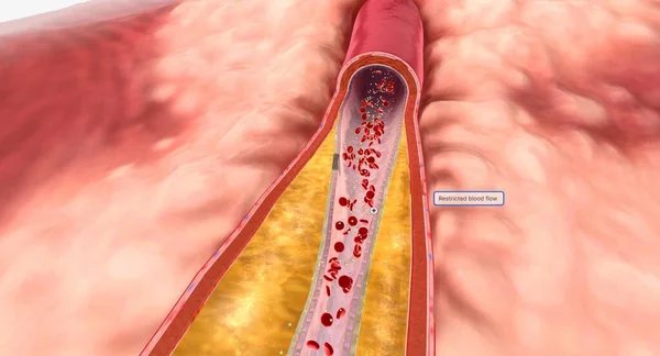
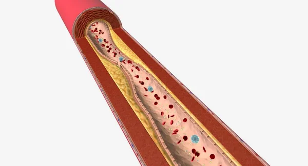
As Hyperlipidemia Continues Over Time, Plaque May Grow And Restrict Blood Flow Through An Artery. 3D Render
Image, 13.87MB, 7258 × 3920 jpg
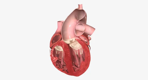
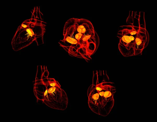
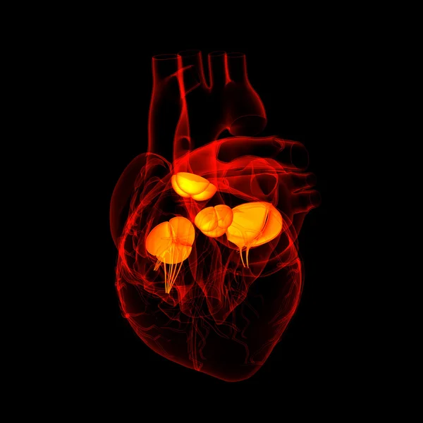
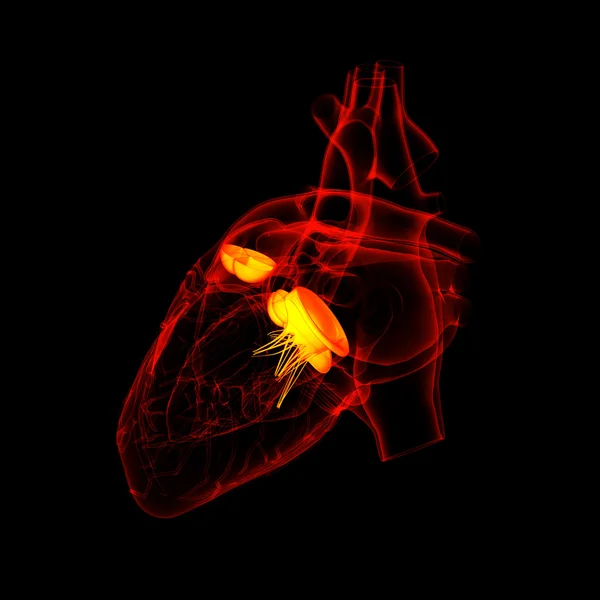
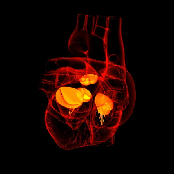
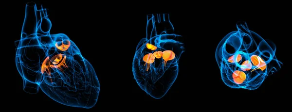
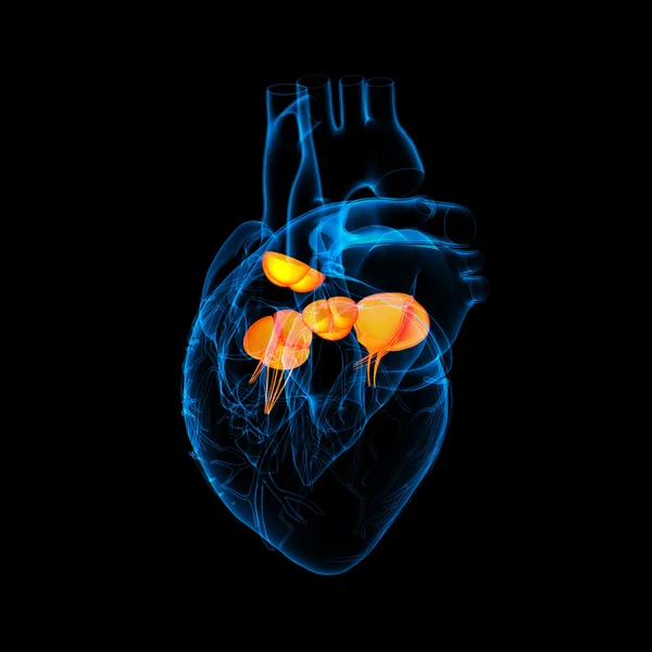
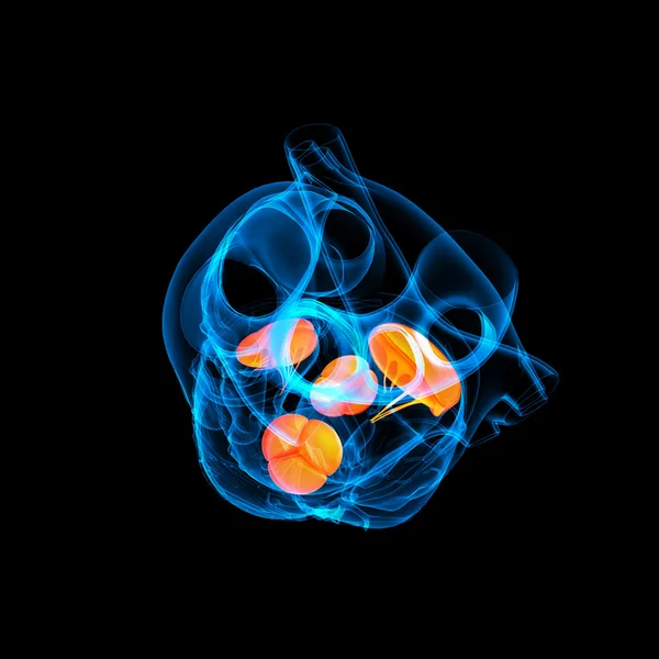
Structure Of The Heart Valves. Mitral Valve, Pulmonary Valve, Aortic Valve And The Tricuspid Valve.
Vector, 9.69MB, 5000 × 3334 eps

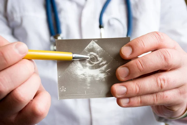
Doctor Holds Snapshot Of Ultrasound Of Heart And Indicates With Ballpoint Pen On Possible Pathology Of Heart Aortic Valve. Concept Photo Of Cardiac Ultrasonic Diagnostics Of Valve Apparatus In Adults
Image, 7.29MB, 6000 × 4000 jpg


Mitral Valve Repair Surgery. Healthy Mitral Valve. Damaged Mitral Valve Before And After Annuloplasty. Vector Illustration
Vector, 7.87MB, 5000 × 3261 eps


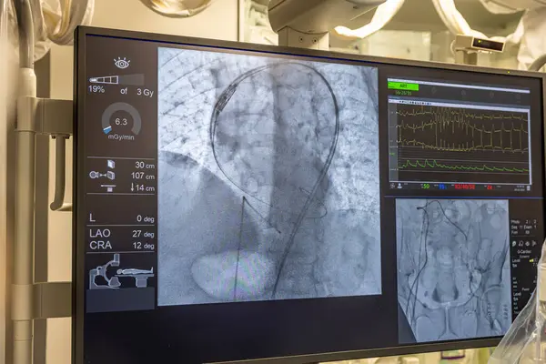
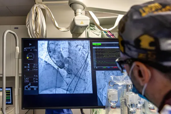
Istanbul, Turkey February 29, 2024; X-ray For Valvular Replacement With Melody Valve. View Of Prosthetic Heart Valve Dysfunction In Patient Who Has Mitral Valve Replacement With Mechanical Valve.
Image, 9.9MB, 5000 × 3335 jpg
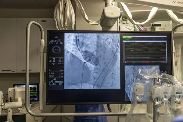
Istanbul, Turkey February 29, 2024; X-ray For Valvular Replacement With Melody Valve. View Of Prosthetic Heart Valve Dysfunction In Patient Who Has Mitral Valve Replacement With Mechanical Valve.
Image, 9.91MB, 5000 × 3335 jpg

Istanbul, Turkey February 29, 2024; X-ray For Valvular Replacement With Melody Valve. View Of Prosthetic Heart Valve Dysfunction In Patient Who Has Mitral Valve Replacement With Mechanical Valve.
Image, 10.13MB, 5000 × 3335 jpg

Istanbul, Turkey February 29, 2024; X-ray For Valvular Replacement With Melody Valve. View Of Prosthetic Heart Valve Dysfunction In Patient Who Has Mitral Valve Replacement With Mechanical Valve.
Image, 10.02MB, 5000 × 3335 jpg

Istanbul, Turkey February 29, 2024; X-ray For Valvular Replacement With Melody Valve. View Of Prosthetic Heart Valve Dysfunction In Patient Who Has Mitral Valve Replacement With Mechanical Valve.
Image, 9.94MB, 5000 × 3335 jpg

Istanbul, Turkey February 29, 2024; X-ray For Valvular Replacement With Melody Valve. View Of Prosthetic Heart Valve Dysfunction In Patient Who Has Mitral Valve Replacement With Mechanical Valve.
Image, 13.81MB, 5000 × 3335 jpg
Istanbul, Turkey February 29, 2024; X-ray For Valvular Replacement With Melody Valve. View Of Prosthetic Heart Valve Dysfunction In Patient Who Has Mitral Valve Replacement With Mechanical Valve.
Image, 9.98MB, 5000 × 3335 jpg

Istanbul, Turkey February 29, 2024; X-ray For Valvular Replacement With Melody Valve. View Of Prosthetic Heart Valve Dysfunction In Patient Who Has Mitral Valve Replacement With Mechanical Valve.
Image, 11.94MB, 5000 × 3335 jpg


Istanbul, Turkey February 29, 2024; X-ray For Valvular Replacement With Melody Valve. View Of Prosthetic Heart Valve Dysfunction In Patient Who Has Mitral Valve Replacement With Mechanical Valve.
Image, 10MB, 5000 × 3335 jpg

Page 1 >> Next