
Stock image C Spine


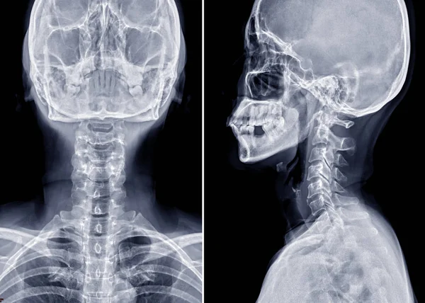
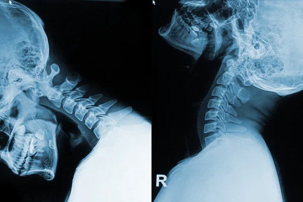
X-ray C-spine Or X-ray Image Of Cervical Spine AP And Lateral View For Diagnostic Intervertebral Disc Herniation.
Image, 2.04MB, 5008 × 2325 jpg
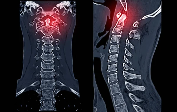

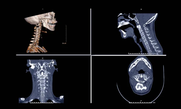
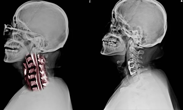
Comparison Of CT C-Spine Or Cervical Spine 3D Rendering Image , Sagittal ,Corona And Axiall View In Patient Trauma Head Injury.
Image, 2.06MB, 5008 × 3008 jpg
X-ray C-spine Or X-ray Image Of Cervical Spine Lateral View For Diagnostic Intervertebral Disc Herniation ,Spondylosis And Fracture.
Image, 2.19MB, 3048 × 3659 jpg
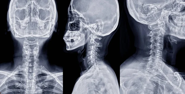
Collection Of X-ray C-spine Or X-ray Image Of Cervical Spine AP , Lateral And Oblique View For Diagnostic Intervertebral Disc Herniation
Image, 14.04MB, 7835 × 4000 jpg
X-ray C-spine Or X-ray Image Of Cervical Spine AP And Lateral View For Diagnostic Intervertebral Disc Herniation
Image, 9.62MB, 5600 × 4000 jpg

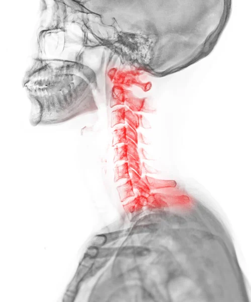
X-ray C-spine Or X-ray Image Of Cervical Spine Lateral View For Diagnostic Intervertebral Disc Herniation. Medical Backgroun
Image, 3.34MB, 3489 × 4000 jpg

X-ray C-spine Or X-ray Image Of Cervical Spine Lateral Extension ,AP And Flexion View For Diagnostic Intervertebral Disc Herniation.
Image, 3.29MB, 6000 × 3306 jpg
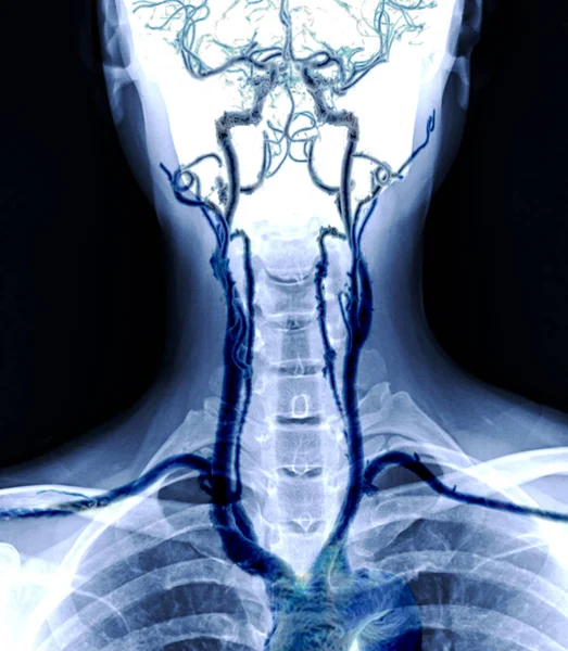
X-ray C-spine Or X-ray Image Of Cervical Spine AP View With Cta Carotid Artery .
Image, 2.73MB, 2713 × 3111 jpg

Collection Chiropractic Logo Design. Spine Logo Template. Spinal Icon. Backbone Icon Related To Physio Therapy
Vector, 0.6MB, 7000 × 3500 eps

C-shaped Scoliosis. Dextroscoliosis. Levoscoliosis. Spinal Curvature, Kyphosis, Lordosis, Scoliosis, Arthrosis. Infographics. Vector Illustration On Isolated Background.
Vector, 8.27MB, 4167 × 4167 eps

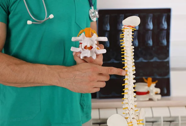
Back Pain Relief Concept. Doctor Chiropractor Explains Causes Of Back Pain Using Spine Model
Image, 16.93MB, 8272 × 5616 jpg
X-ray Of The Left Collarbone. Fracture Of Clavicle Of The Child. Consolidation Of The Fracture. Positive. Negative.
Image, 3.56MB, 4014 × 2429 jpg
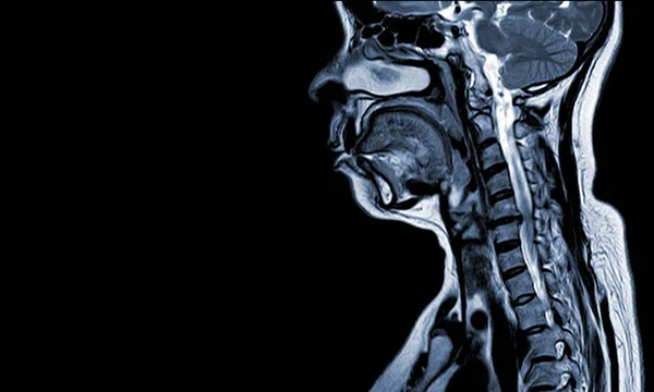
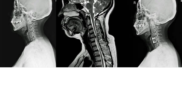
A Sagittal View Of MRI C-spine Or Magnetic Resonance Image Of Cervical Spine Showing Spondylosis Causing Cervical Spondylotic Myelopathy And Compression Fracture.
Image, 1.46MB, 2835 × 2976 jpg
MRI C-spine Moderate Spinal Cord Compression At C5-6, Medical Image Concept, And Copy Space.
Image, 4.46MB, 7000 × 4205 jpg

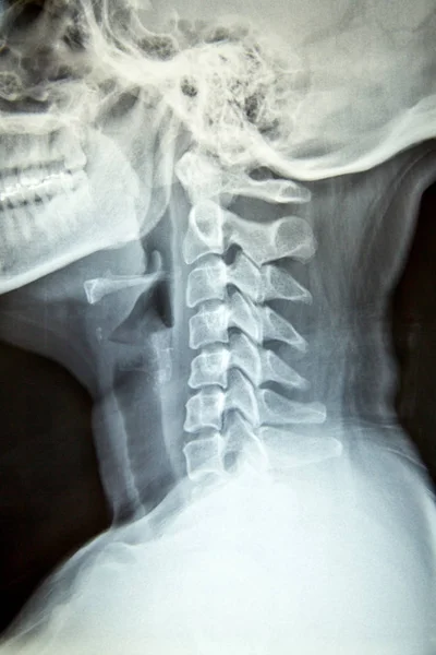
Close Up X-ray Film Show Cervical Spine Or C-spine, Neck Bones X-ray Film. Radiology To Educate Human Anatomy. Office Syndrome And Cervical Spondylosis Concept.
Image, 12.97MB, 5760 × 3840 jpg

Close Up X-ray Film Show Cervical Spine Or C-spine, Neck Bones X-ray Film. Radiology To Educate Human Anatomy. Office Syndrome And Cervical Spondylosis Concept.
Image, 11.64MB, 3840 × 5760 jpg
Close Up X-ray Film Show Cervical Spine Or C-spine, Neck Bones X-ray Film. Radiology To Educate Human Anatomy. Office Syndrome And Cervical Spondylosis Concept.
Image, 10.91MB, 3840 × 5760 jpg
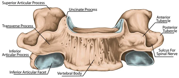
Didactic Board, Cervical Spine, Common Vertebral Morphology, Sixth Cervical Vertebra, Cervical Vertebrae, Anterior View
Image, 3.37MB, 5906 × 2588 jpg
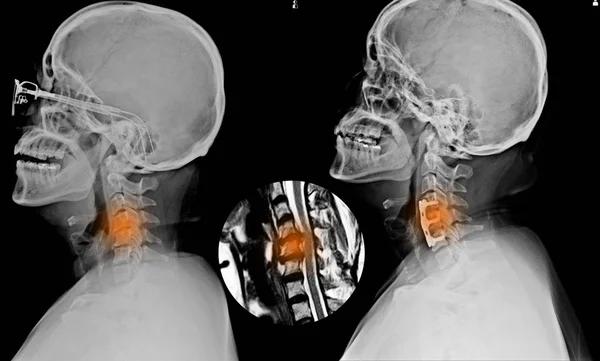
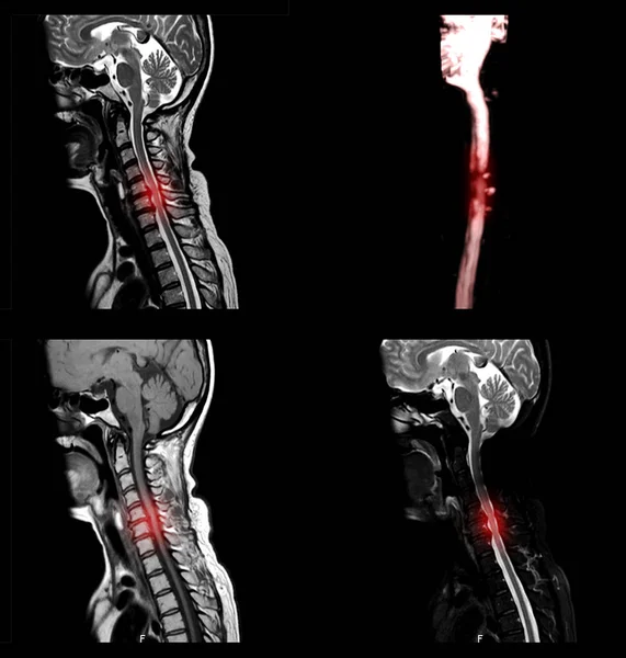
X-ray C-SPINES Showing S/p Internal Fixation C4-C5 & C6 With Plate & Screws And There Is Hypersignal Intensity Lesion In The Spinal Cord At C4 To C6 Levels.
Image, 4.46MB, 4972 × 3000 jpg
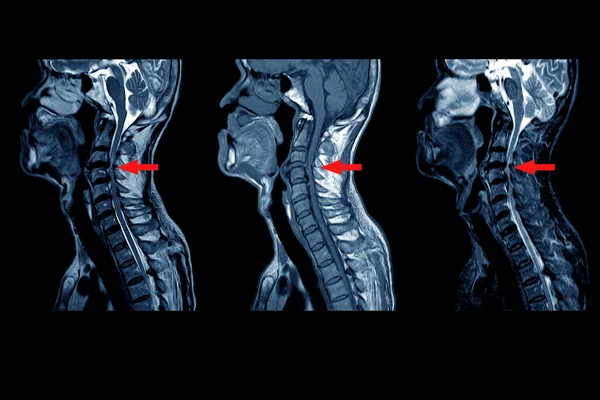
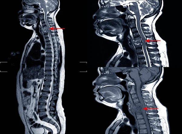
MRI OF CERVICAL SPINE : Moderate To Severe Posterior Central Disc Protrusion Of C3/4 To C5/6 Intervertebral Discs With A 2.0 Cm In Length Small Posterior Subligamentous Fluid Collection.on Red Point
Image, 4.36MB, 6000 × 4000 jpg
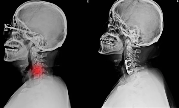
X-ray C-SPINES Showing S/p Internal Fixation C4-C5 & C6 With Plate & Screws And There Is Hypersignal Intensity Lesion In The Spinal Cord At C4 To C6 Levels.
Image, 4.38MB, 4972 × 3000 jpg
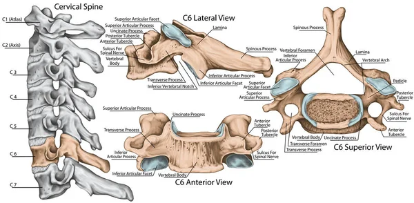
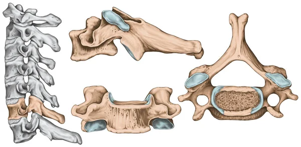
Didactic Board, Cervical Spine, Common Vertebral Morphology, Sixth Cervical Vertebra, Cervical Vertebrae, Anterior, Lateral And Superior View
Image, 4.61MB, 5906 × 2902 jpg

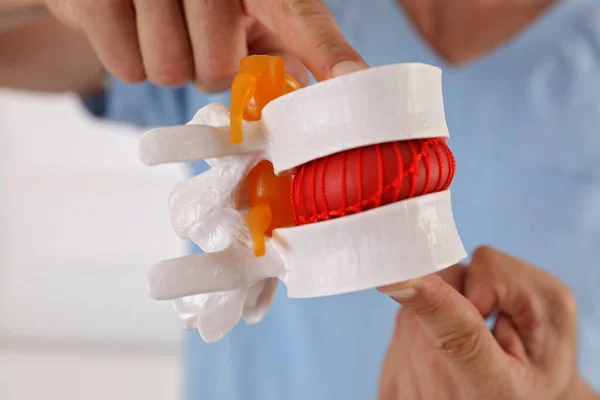
Back Pain Relief Concept. Doctor Chiropractor Explains Causes Of Back Pain Lumbar Spine Disk Model
Image, 18.96MB, 8656 × 5696 jpg

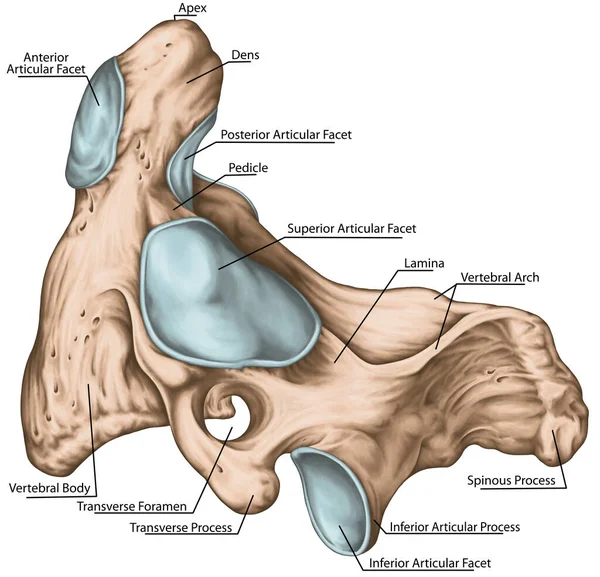
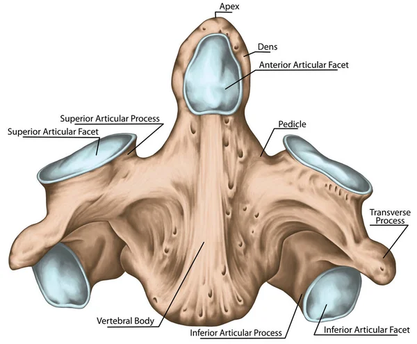
Didactic Board, Cervical Spine, Vertebral Morphology, Second Cervical Vertebra, Axis, Cervical Vertebrae, Transverse Foramen, Vertebral Foramen, Dens, Odontoid, Anterior And Posterior Articular Facet, Lateral View
Image, 6.33MB, 5906 × 5688 jpg
Didactic Board, Cervical Spine, Common Vertebral Morphology, Sixth Cervical Vertebra, Cervical Vertebrae, Anterior, Lateral And Superior View
Image, 3.91MB, 5906 × 2902 jpg
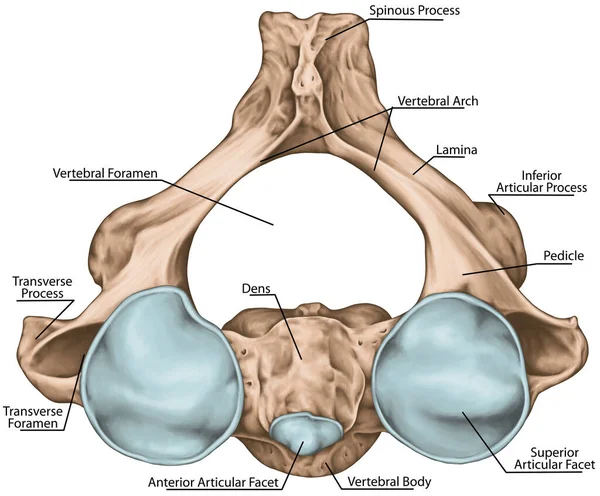
Didactic Board, Cervical Spine, Vertebral Morphology, Second Cervical Vertebra, Axis, Cervical Vertebrae, Dens, Odontoid, Anterior View
Image, 5.47MB, 5906 × 4880 jpg
Back Pain Relief Concept. Doctor Chiropractor Explains Causes Of Back Pain Lumbar Spine Disk Model
Image, 16.19MB, 8688 × 5792 jpg
X-ray C-SPINES Showing S/p Internal Fixation C4-C5 & C6 With Plate & Screws And There Is Hypersignal Intensity Lesion In The Spinal Cord At C4 To C6 Levels.
Image, 3.41MB, 4972 × 3000 jpg

Cervical Spine Spondylosis, X-ray C-spine Show Degenerative Change And Loss Space Of Nerve Root Foramen
Image, 2.34MB, 2936 × 1771 jpg


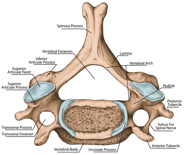
Didactic Board, Cervical Spine, Common Vertebral Morphology, Sixth Cervical Vertebra, Cervical Vertebrae, Transverse Foramen, Vertebral Foramen, Superior View
Image, 5.48MB, 5906 × 4962 jpg
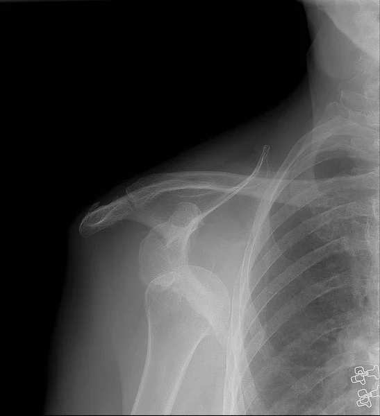
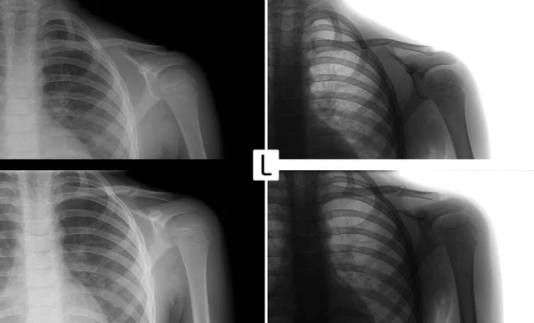
X-ray Image Of Shoulder, Shows Right Shoulder Dislocation. Anterior Projection
Image, 1.35MB, 2028 × 2224 jpg

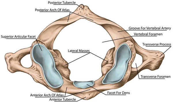
Didactic Board, Cervical Spine, Vertebral Morphology, First Cervical Vertebra, Atlas, Cervical Vertebrae, Transverse Foramen, Vertebral Foramen, Facet For Dens, Superior View
Image, 3.88MB, 5906 × 3517 jpg
Didactic Board, Cervical Spine, Vertebral Morphology, Second Cervical Vertebra, Axis, Cervical Vertebrae, Transverse Foramen, Vertebral Foramen, Dens, Odontoid, Anterior And Posterior Articular Facet, Superior View
Image, 5.47MB, 5906 × 4884 jpg
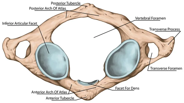
Didactic Board, Cervical Spine, Vertebral Morphology, First Cervical Vertebra, Atlas, Cervical Vertebrae, Transverse Foramen, Vertebral Foramen, Facet For Dens, Inferior View
Image, 3.57MB, 5906 × 3340 jpg
Page 1 >> Next