
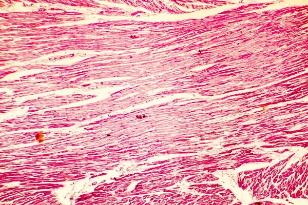
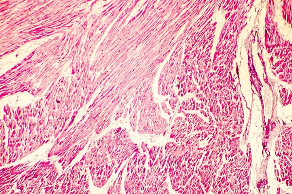
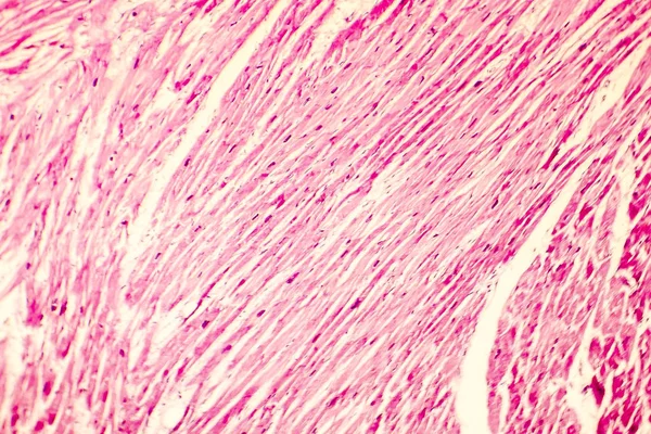
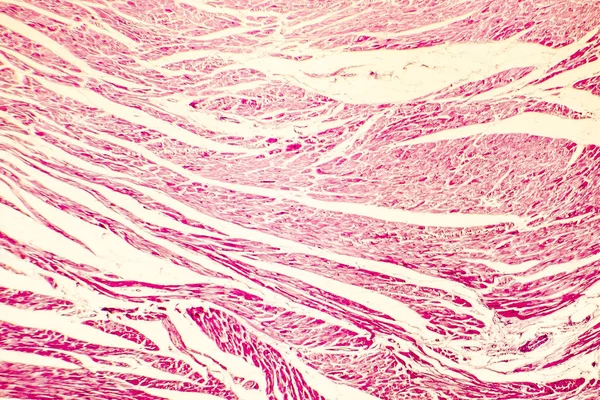
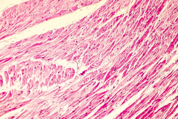
Stock image Cardiac Hypertrophy
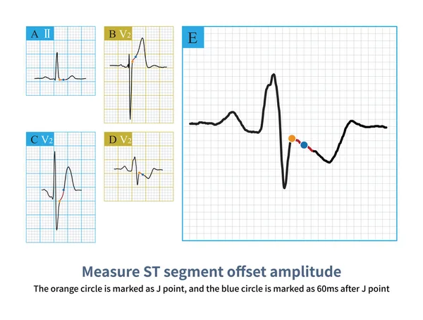
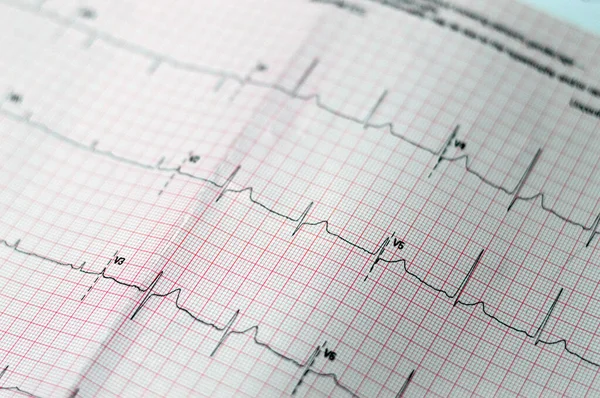
Firstly, Select Point J As The Reference Point, And Then Select 60ms After Point J As The Measurement Point To Evaluate The ST Segment Offset Morphology And Amplitude.
Image, 10.33MB, 10000 × 7579 jpg
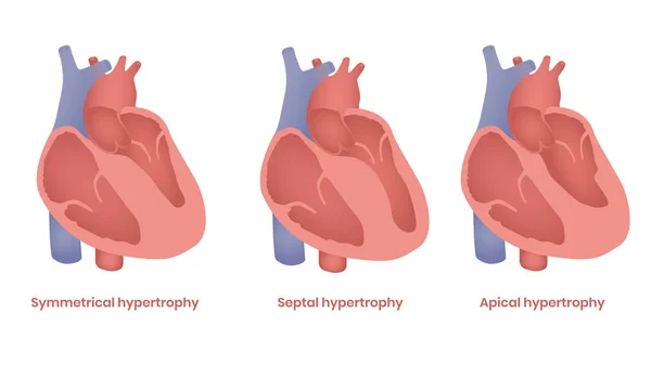
Hypertrophic Cardiomyopathy Illustration. Apical, Septal And Symmetrical Types
Vector, 4.68MB, 7111 × 4000 eps
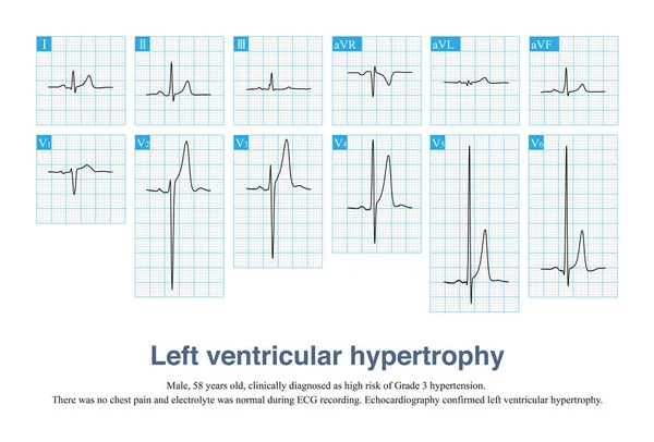
Sometimes, Left Ventricular Hypertrophy With Tall T Waves Is Easily Misdiagnosed As Hyperkalemia And Hyperacute T Waves, And ECG Needs To Be Carefully Identified In Combination With Clinic.
Image, 13.77MB, 10000 × 6782 jpg
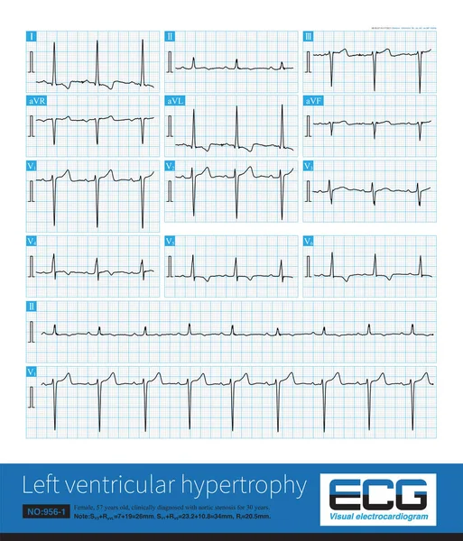
Sometimes, Because The QRS Axis Is In The Upper Left Quadrant, The High-amplitude R Wave Of Left Ventricular Hypertrophy Occurs In The Limb Leads, And Left Chest Leads Is Normal.
Image, 31.4MB, 10000 × 11694 jpg

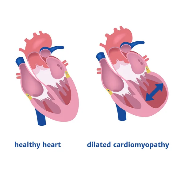
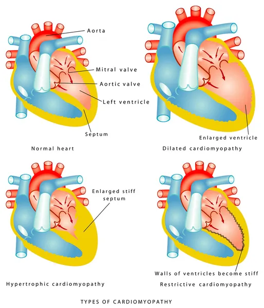
Dilated Cardiomyopathy. Expansion Of The Ventricle Of The Heart. Medical Poster Vector Illustration
Vector, 5.93MB, 2000 × 2000 eps
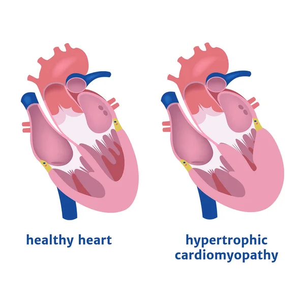
Hypertrophic Cardiomyopathy. Expansion Of The Ventricle Of The Heart. Medical Poster Vector Illustration
Vector, 6.01MB, 2000 × 2000 eps

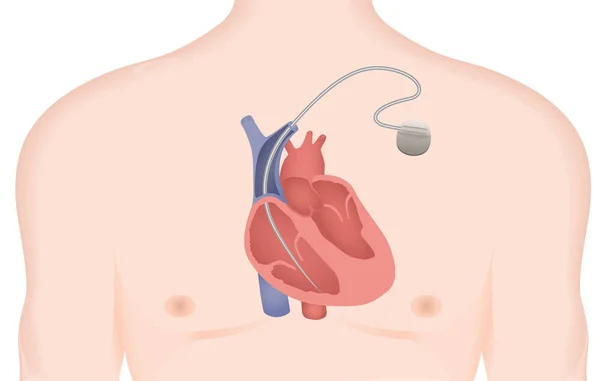
Artificial Cardiac Pacemaker Vector Illustration. Implantable Cardioverter Defibrillator
Vector, 8.24MB, 6289 × 4000 eps
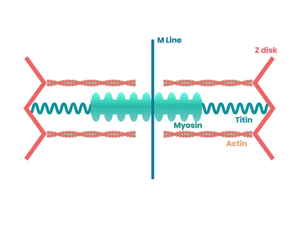
Heart Muscle Proteins And Philament Structure. Myosin, Actin And Titin Illustration
Vector, 2.39MB, 5156 × 4000 eps
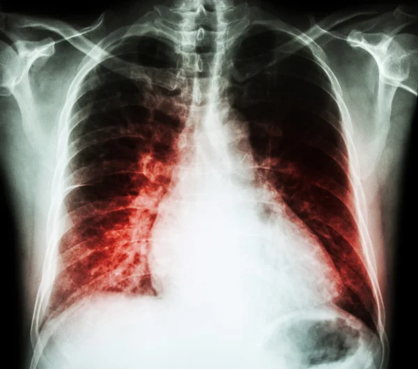
Heart Failure ( Film X-ray Chest PA Upright : Show Cardiomegaly And Interstitial Infiltrate Both Lung )
Image, 5.88MB, 3915 × 3456 jpg

Mitral Valve Repair Surgery. Healthy Mitral Valve. Damaged Mitral Valve Before And After Annuloplasty. Vector Illustration
Vector, 7.87MB, 5000 × 3261 eps


Research Scientist. Science Laboratory, Chemistry Scientists And Clinical Lab. Medical Research Items, Clinical Science Laboratories Experiments. Heart, Cardiac Problem, Concept Vector Illustration
Vector, 8.55MB, 8333 × 5001 eps

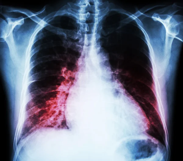
Heart Failure ( Film X-ray Chest PA Upright : Show Cardiomegaly And Interstitial Infiltrate Both Lung )
Image, 6.45MB, 3915 × 3456 jpg

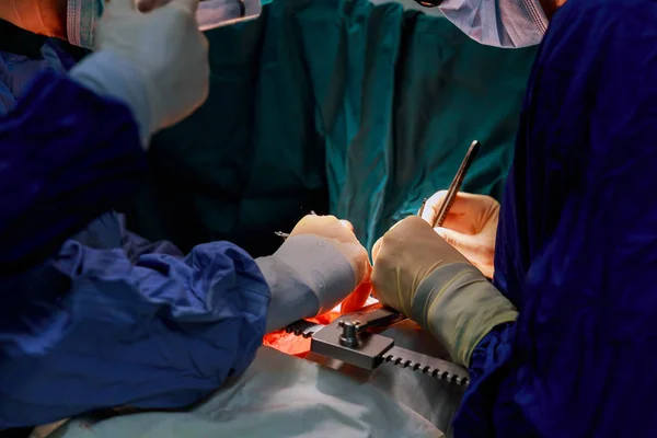
Doctor Doing Heart Operation Donor Heart Procurement For Cardiac Transplantation
Image, 7.45MB, 4500 × 3000 jpg


Cardiomyopathy - Diagnosis Written On A White Piece Of Paper. Syringe And Vaccine With Drugs.
Image, 10.88MB, 5184 × 3456 jpg
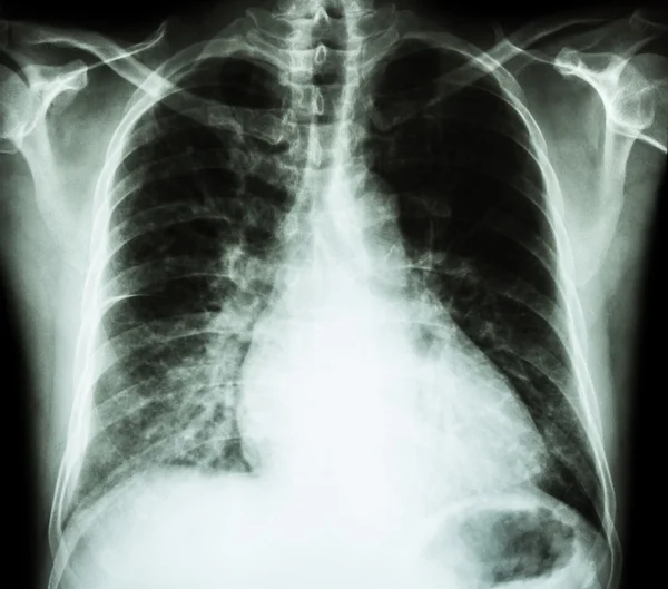
Heart Failure ( Film X-ray Chest PA Upright : Show Cardiomegaly And Interstitial Infiltrate Both Lung )
Image, 4.93MB, 3915 × 3456 jpg

Heart Failure ( Film X-ray Chest PA Upright : Show Cardiomegaly And Interstitial Infiltrate Both Lung )
Image, 6.71MB, 3915 × 3456 jpg


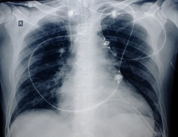
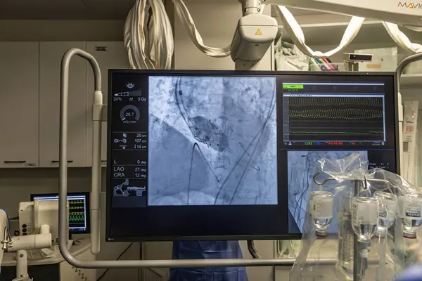
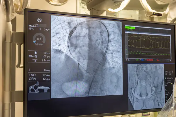
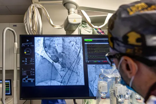
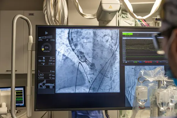
Istanbul, Turkey February 29, 2024; X-ray For Valvular Replacement With Melody Valve. View Of Prosthetic Heart Valve Dysfunction In Patient Who Has Mitral Valve Replacement With Mechanical Valve.
Image, 9.9MB, 5000 × 3335 jpg
Istanbul, Turkey February 29, 2024; X-ray For Valvular Replacement With Melody Valve. View Of Prosthetic Heart Valve Dysfunction In Patient Who Has Mitral Valve Replacement With Mechanical Valve.
Image, 9.91MB, 5000 × 3335 jpg

Istanbul, Turkey February 29, 2024; X-ray For Valvular Replacement With Melody Valve. View Of Prosthetic Heart Valve Dysfunction In Patient Who Has Mitral Valve Replacement With Mechanical Valve.
Image, 10.13MB, 5000 × 3335 jpg

Istanbul, Turkey February 29, 2024; X-ray For Valvular Replacement With Melody Valve. View Of Prosthetic Heart Valve Dysfunction In Patient Who Has Mitral Valve Replacement With Mechanical Valve.
Image, 10.02MB, 5000 × 3335 jpg

Istanbul, Turkey February 29, 2024; X-ray For Valvular Replacement With Melody Valve. View Of Prosthetic Heart Valve Dysfunction In Patient Who Has Mitral Valve Replacement With Mechanical Valve.
Image, 9.94MB, 5000 × 3335 jpg

Istanbul, Turkey February 29, 2024; X-ray For Valvular Replacement With Melody Valve. View Of Prosthetic Heart Valve Dysfunction In Patient Who Has Mitral Valve Replacement With Mechanical Valve.
Image, 13.81MB, 5000 × 3335 jpg
Istanbul, Turkey February 29, 2024; X-ray For Valvular Replacement With Melody Valve. View Of Prosthetic Heart Valve Dysfunction In Patient Who Has Mitral Valve Replacement With Mechanical Valve.
Image, 9.98MB, 5000 × 3335 jpg
Istanbul, Turkey February 29, 2024; X-ray For Valvular Replacement With Melody Valve. View Of Prosthetic Heart Valve Dysfunction In Patient Who Has Mitral Valve Replacement With Mechanical Valve.
Image, 11.94MB, 5000 × 3335 jpg
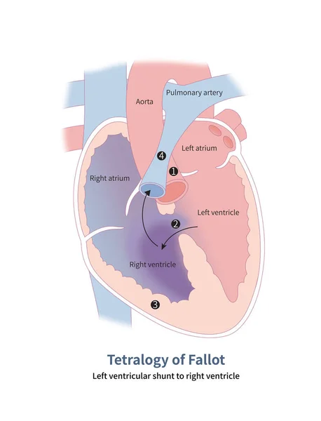
Four Anatomical Malformations Of Tetralogy Of Fallot: 1 Aortic Straddling; 2 Ventricular Septal Defect; 3 Right Ventricular Hypertrophy And 4 Pulmonary Artery Stenosis.
Image, 12.08MB, 10000 × 13070 jpg

Istanbul, Turkey February 29, 2024; X-ray For Valvular Replacement With Melody Valve. View Of Prosthetic Heart Valve Dysfunction In Patient Who Has Mitral Valve Replacement With Mechanical Valve.
Image, 10MB, 5000 × 3335 jpg
Istanbul, Turkey February 29, 2024; X-ray For Valvular Replacement With Melody Valve. View Of Prosthetic Heart Valve Dysfunction In Patient Who Has Mitral Valve Replacement With Mechanical Valve.
Image, 10.38MB, 5000 × 3335 jpg
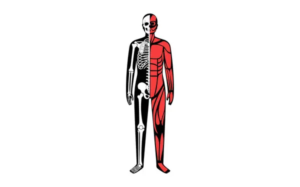
Skeleton With Muscles, Joints, Cartilages And Ligaments In The Human Body. Muscle Fibers And Connective Tissue Sheaths In The Male Silhouette. Musculoskeletal Anatomy Flat Vector Medical Illustration
Vector, 0.53MB, 5694 × 3551 eps

Istanbul, Turkey February 29, 2024; X-ray For Valvular Replacement With Melody Valve. View Of Prosthetic Heart Valve Dysfunction In Patient Who Has Mitral Valve Replacement With Mechanical Valve.
Image, 9.94MB, 5000 × 3335 jpg
ECG ElectroCardioGraph Paper That Shows Sinus Rhythm Abnormality Of Right Ventricular Hypertrophy, Inferior T Wave Due To Hypertrophy And Ischemia, Abnormal ECG Study, Unconfirmed Diagnosis
Image, 17.21MB, 6016 × 4000 jpg

Istanbul, Turkey February 29, 2024; X-ray For Valvular Replacement With Melody Valve. View Of Prosthetic Heart Valve Dysfunction In Patient Who Has Mitral Valve Replacement With Mechanical Valve.
Image, 10.4MB, 5000 × 3335 jpg

Istanbul, Turkey February 29, 2024; X-ray For Valvular Replacement With Melody Valve. View Of Prosthetic Heart Valve Dysfunction In Patient Who Has Mitral Valve Replacement With Mechanical Valve.
Image, 9.98MB, 5000 × 3335 jpg

Istanbul, Turkey February 29, 2024; X-ray For Valvular Replacement With Melody Valve. View Of Prosthetic Heart Valve Dysfunction In Patient Who Has Mitral Valve Replacement With Mechanical Valve.
Image, 9.91MB, 5000 × 3335 jpg

Istanbul, Turkey February 29, 2024; X-ray For Valvular Replacement With Melody Valve. View Of Prosthetic Heart Valve Dysfunction In Patient Who Has Mitral Valve Replacement With Mechanical Valve.
Image, 9.92MB, 5000 × 3335 jpg
Page 1 >> Next