
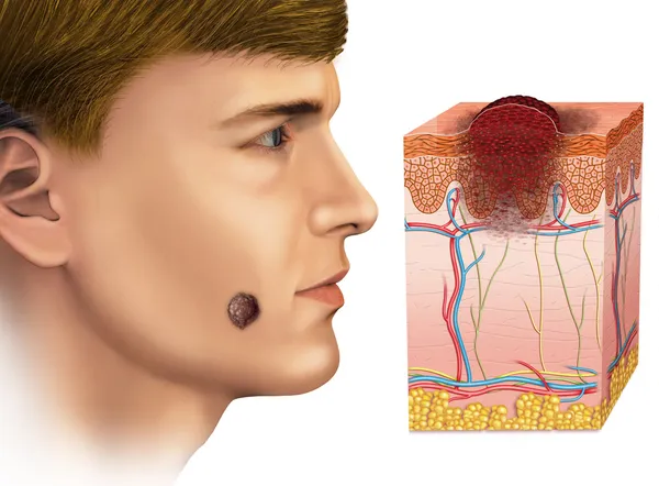
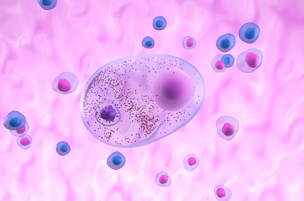
Stock image Cell Degeneration
Brain, Neurons, Synapses, Neural Network Circuit Of Neurons, Degenerative Diseases, Parkinson, 3d Rendering
Image, 14.15MB, 5177 × 3897 jpg
Lewy Bodies In Parkinson's Disease (PD) Or Dementia (LBD) - Isometric 3d Illustration
Image, 5.18MB, 10000 × 6600 jpg
Lewy Body In Parkinson's Disease (PD) Or Dementia (LBD) - Closeup 3d Illustration
Image, 4.62MB, 10000 × 6600 jpg
Lewy Body In Parkinson's Disease (PD) Or Dementia (LBD) - Isometric 3d Illustration
Image, 3.65MB, 10000 × 6600 jpg
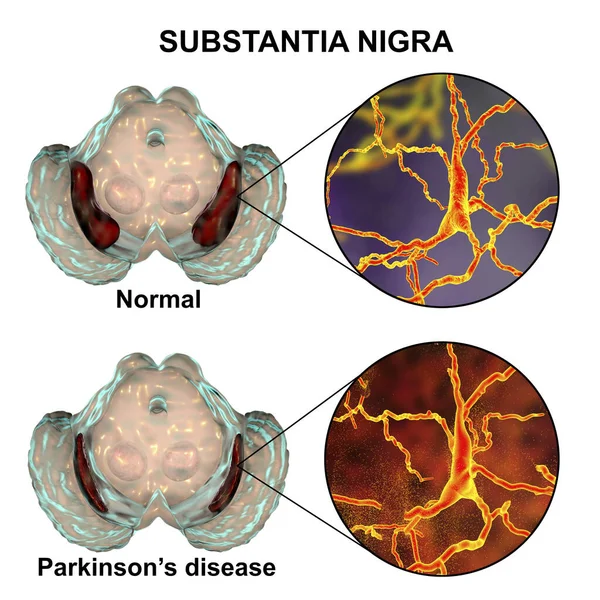
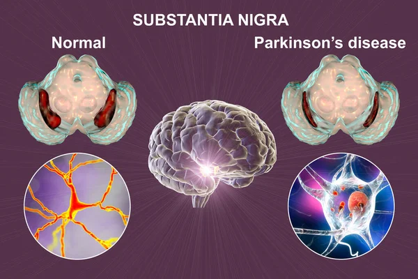
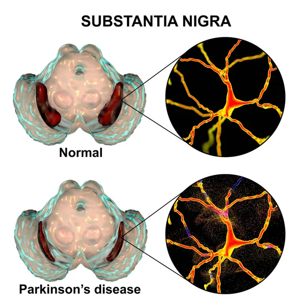
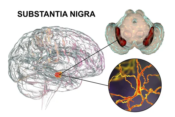
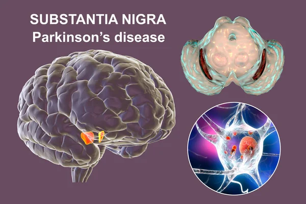
Substantia Nigra In Norm And In Parkinson's Disease, 3D Illustration Showing Decrease Of Its Volume. There Is Degeneration Of Dopaminergic Neurons In The Pars Compacta Of The Substantia Nigra
Image, 4.57MB, 6000 × 6000 jpg

Black Substance Of The Midbrain And Its Dopaminergic Neurons In Normal State And In Parkinson's Disease. 3D Illustration Showing Volume Decrease And Accumulation Of Lewy Bodies In Neurons
Image, 20.15MB, 9200 × 6133 jpg
Substantia Nigra In Norm And In Parkinson's Disease, 3D Illustration Showing Decrease Of Its Volume. There Is Degeneration Of Dopaminergic Neurons In The Pars Compacta Of The Substantia Nigra
Image, 4.35MB, 6000 × 6000 jpg
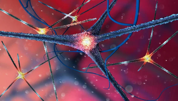
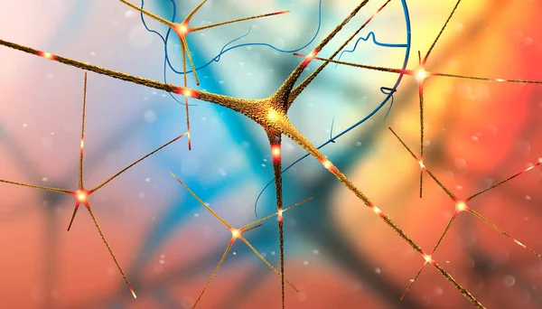
Microscopic View Of The Synapses. Brain Connections. Neurons And Synapses. Communication And Cerebral Stimulus. Neural Network Circuit, Degenerative Diseases, Parkinson, Alzheimer. 3d Render
Image, 10.63MB, 5511 × 3149 jpg

Microscopic View Of The Synapses. Brain Connections. Neurons And Synapses. Communication And Cerebral Stimulus. Neural Network Circuit, Degenerative Diseases, Parkinson, Alzheimer. 3d Render
Image, 7.98MB, 5511 × 3149 jpg

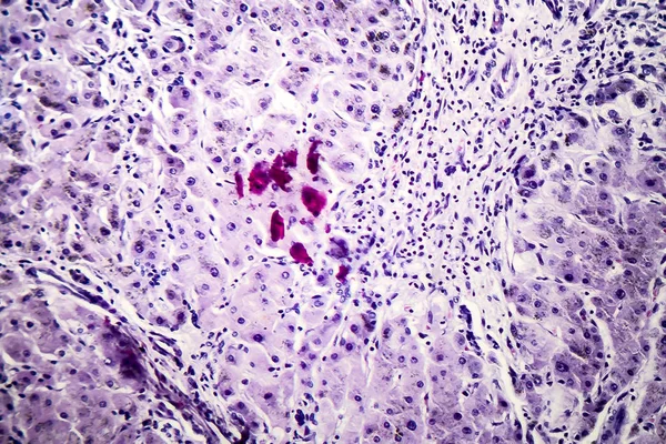
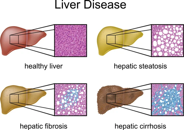
Liver With Cirrhosis Inside Human Body. 3D Illustration And Light Micrograph Of Small Nodular Cirrhosis
Image, 9.28MB, 5570 × 3133 jpg

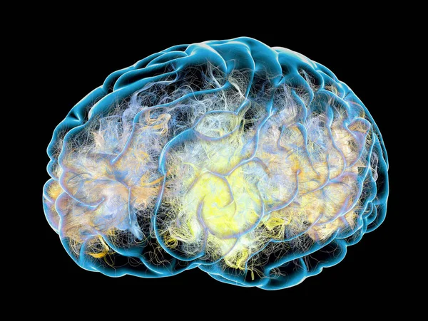
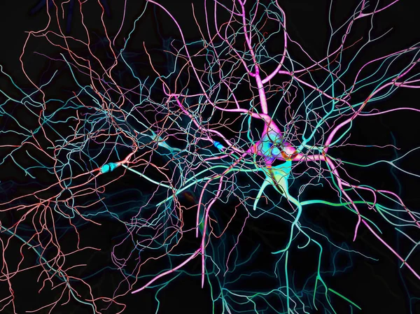
Neurons, Highly Detailed Brain Cells, Neural Network, 3D Illustration
Image, 12.08MB, 5120 × 5120 jpg
Caudate Nuclei In Human Brain And Its Neurons, 3D Illustration. The Caudate Nucleus Is A Component Of The Basal Ganglia, It Plays Role In Choreas, Neurodegenerative And Other Brain Diseases
Image, 16.77MB, 8157 × 5438 jpg
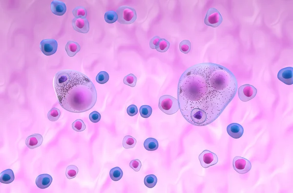
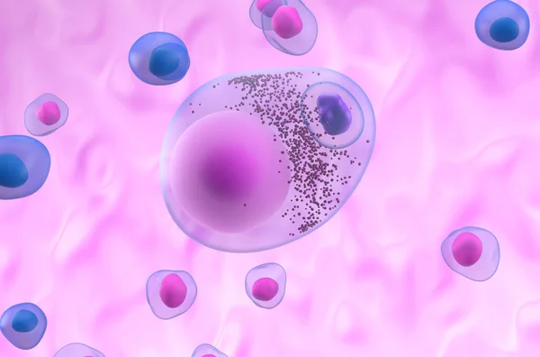
A Microglia Cell. It Plays An Important Role In The Pathogenesis Of Alzheimer's Disease
Image, 13.48MB, 8000 × 6000 jpg
Microscopic View Of The Synapses. Brain Connections. Neurons And Synapses. Communication And Cerebral Stimulus. Neural Network Circuit, Degenerative Diseases, Parkinson, Alzheimer. 3d Render
Image, 9.46MB, 5511 × 3149 jpg

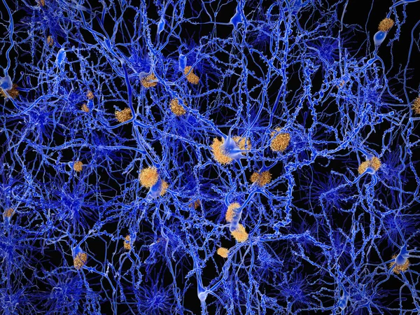
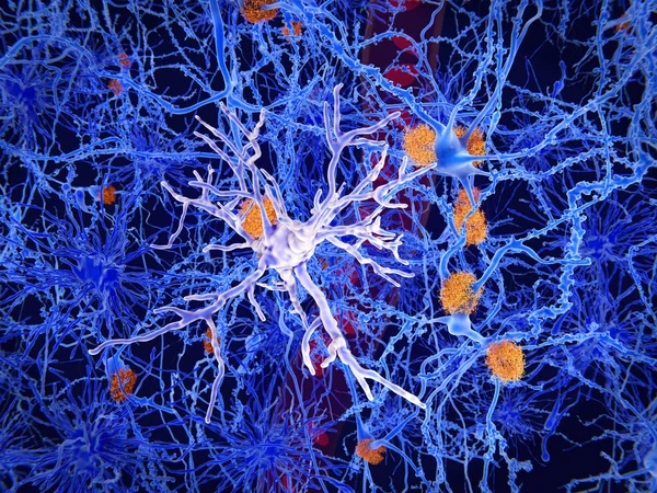
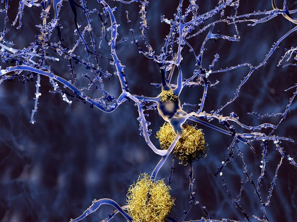
Alzheimer Disease, Neuron Network With Amyloid Plaques. Illustration
Image, 28.11MB, 8000 × 6000 jpg
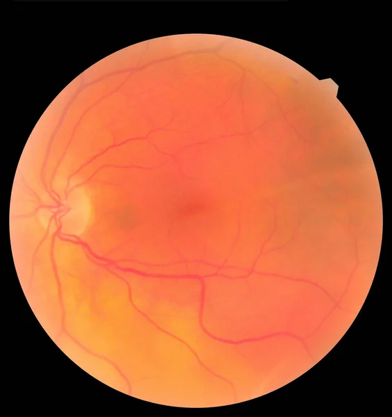
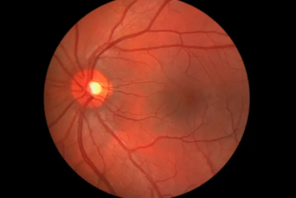
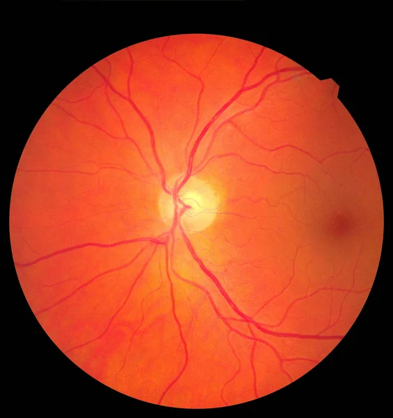
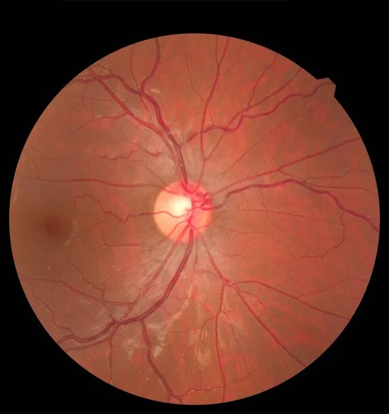
Ophthalmic Image Detailing The Retina And Optic Nerve Inside A Healthy Human Eye. Health Protection Concept
Image, 2.77MB, 3000 × 3186 jpg

Caudate Nuclei In Human Brain And Its Neurons, 3D Illustration. The Caudate Nucleus Is A Component Of The Basal Ganglia, It Plays Role In Choreas, Neurodegenerative And Other Brain Diseases
Image, 12.8MB, 8157 × 5438 jpg

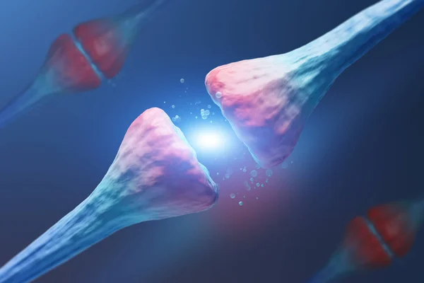
Neural Synapses, Failure In Their Functioning Causes Degenerative Neurological Diseases Such As Alzheimer's, Parkson's And Dementia. 3D Rendering
Image, 2.96MB, 3508 × 2480 jpg

Destruction Of Neurons Of The Caudate Nucleus, Conceptual 3D Illustration. Caudate Nucleus Belongs To The Brain Basal Ganglia, Its Neurons Are Damaged In Huntingon's Disease And Other Choreas
Image, 16.49MB, 7996 × 5331 jpg
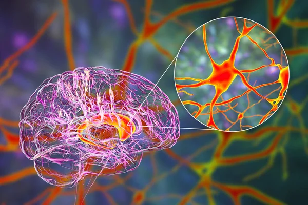
Black Substance Of The Midbrain And Its Dopaminergic Neurons, 3D Illustration. Black Substance Regulates Movement And Reward, Its Degeneration Is A Key Step In Development Of Parkinson's Disease
Image, 11.11MB, 7711 × 5140 jpg
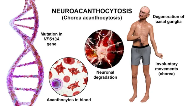
Neuroacanthocytosis, Chorea Acanthocytosis, A Neurodegenerative Disease Due To Mutation In The Gene VPS13A, It Is Marked By Presence Of Acanthocytes In Blood And Choreiform Movements, 3D Illustration
Image, 13.25MB, 9117 × 5128 jpg
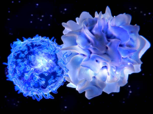
Interaction Between A Dendritic Cell And A T-lymphocyte. 3d-rendering. Dendritic Cells Are Antigen-presenting Cells Of The Immune System
Image, 6.52MB, 8000 × 6000 jpg


Infectious Etiology Of Dementia. Neuropsychiatric Sequelae Of Covid-19. Viruses Infecting Neurons And Progressive Impairment Of Brain Functions, Amyloid Plaques In Brain Tissues. 3D Illustration
Image, 14.68MB, 7200 × 4050 jpg

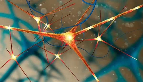
Brain Neurons Synapse Functions. Neural Network Circuit Of Neurons, Degenerative Diseases, Parkinson
Image, 1.6MB, 4724 × 3937 jpg

Ophthalmic Image Detailing The Retina And Optic Nerve Inside A Healthy Human Eye. Health Protection Concept
Image, 2.67MB, 3000 × 3186 jpg

Ophthalmic Image Detailing The Retina And Optic Nerve Inside A Healthy Human Eye. Medicine Concept
Image, 3.76MB, 3000 × 3186 jpg
Abstract Image Of Gray And Pink Diagonal Neurons Over Dark Blue Background With Nervous Cells. Concept Of Science And Medicine. 3d Rendering
Image, 1.17MB, 4500 × 3000 jpg
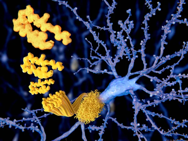
The Beta Amyloid Peptid, Amyloid Plaques Growing On A Neuron. It Consists Of About 30 Amino Acids And Aggregates To Amyloid Plaques, That May Damage And Kill Neurons. Illustration
Image, 4.49MB, 8000 × 6000 jpg
Brain, Neurons, Synapses, Neural Network Circuit Of Neurons, Degenerative Diseases, Parkinson
Image, 15.81MB, 5511 × 4133 jpg
Ophthalmic Image Detailing The Retina And Optic Nerve Inside A Healthy Human Eye. Medicine Concept
Image, 3.41MB, 3000 × 3186 jpg

Ophthalmic Image Detailing The Retina And Optic Nerve Inside A Healthy Human Eye. Health Protection Concept
Image, 2.69MB, 3000 × 3186 jpg
Black Substance Of The Midbrain In Parkinson's Disease, 3D Illustration Showing Decrease Of Its Volume And Accumulation Of Lewy Bodies In Dopaminergic Neurons Of The Black Substance
Image, 8.32MB, 7711 × 5140 jpg

Abstract Image Of White Diagonal Neurons Over Gray Background With Nervous Cells. Concept Of Science And Medicine. 3d Rendering
Image, 1.26MB, 4500 × 3000 jpg


Eye Disease, Atrophic Age-Related Macular Degeneration Illustrated - Translation: Normal State, Atrophic Age-related Macular Degeneration, Atrophy Of Macular Tissue With Age, Retina, Choroid, Central Fossa (macula)
Vector, 0.25MB, 5334 × 3334 ai

Page 1 >> Next