
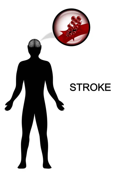
Stock image Cerebrovascular page 3

Blurry Hospital Hallway With Result Of Human Head MRI Scan On The Computer Display, Unfocused Background.
Image, 3.24MB, 4000 × 2667 jpg

Man Suffering Of A Cerebrovascular Accident Or Stroke Or Brain Attack With Blood Clot Or Thrombus 3D Rendering Illustration. Medicine, Medical Pathology, Health, Brain Injury, Science Concepts.
Image, 3.34MB, 3500 × 2000 jpg

CT Scan Of The Brain Sagittal View For Diagnosis Brain Tumor,stroke Diseases And Vascular Diseases.
Image, 1.34MB, 3380 × 2862 jpg
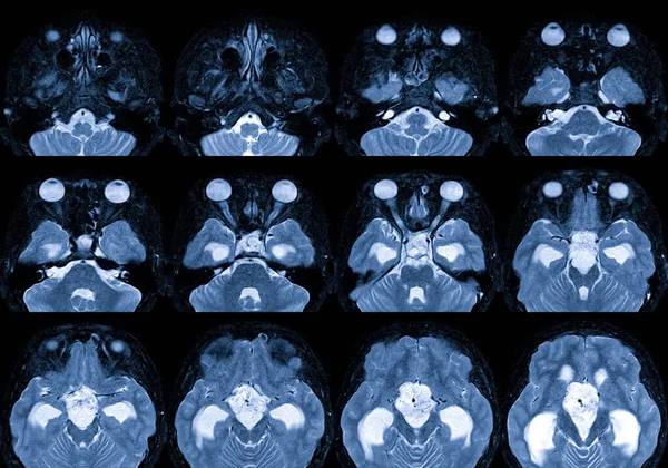
MRA AND MRV OF BRAIN Finding:Bilateral Territorial Muscles To The Cortex And Subcortical Of The Parietal And Low Hemispheres Cerebellar.
Image, 7.85MB, 4540 × 2430 jpg

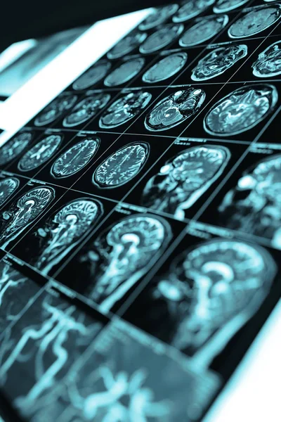
Selective Focus Of MRI Brain Sagittal Plane For Detect A Variety Of Conditions Of The Brain Such As Cysts, Tumors, Bleeding, Swelling, Developmental And Structural Abnormalities Or Infections .
Image, 6.81MB, 7056 × 5208 jpg
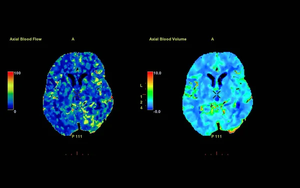
Perfusion CT Scan Of The Brain ,The Red And Green And Blue Are Areas Of Delayed Blood Flow To The Brain On The Perfusion CT Scan On The Screen.
Image, 1.89MB, 3933 × 2476 jpg
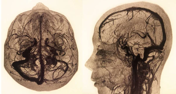
Cerebral Angiography Image From Fluoroscopy In Intervention Radiology Showing Cerebral Artery.
Image, 5.75MB, 6000 × 4000 jpg

Axial View Of CT Perusion Of The Brain With Contrast Showing Brain Anatomy, Lobes, Perfusion And Function. The Red Area Indicates High Brain Activity, Perfusion And Function.
Image, 2.32MB, 2814 × 3096 jpg
MRI Brain With Contrast Media Finding: There Is A 3.5cm Diameter Lobulated Mass At Suprasellar With Compression Of The Pituitary Gland,Medical Healthcare Concept.
Image, 7.79MB, 6000 × 4204 jpg
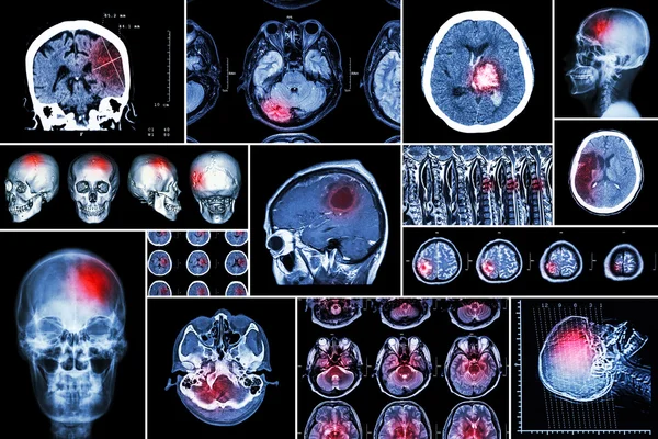
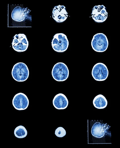
Set , Collection Of Brain Disease ( Cerebral Infarction , Hemorrhagic Stroke , Brain Tumor , Disc Herniation With Spinal Cord Compression ,etc)( CT Scan , MRI , MRT )( Neurology And Nervous System )
Image, 12.02MB, 6000 × 4000 jpg

Magnetic Resonance Imaging Of The Head Of A Healthy Person, Image Of The Pituitary Gland
Image, 11.51MB, 6240 × 4160 jpg

Cerebral Angiography Is A Specialized Procedure To Visualize The Arteries And Veins In The Brain For Medical Diagnosis.
Image, 1.89MB, 3072 × 3072 jpg

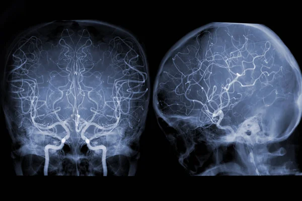
MRA Brain And Neck Or Magnetic Resonance Angiography ( MRA ) Of Cerebral Artery And Common Carotid Artery For Evaluate Them Stenosis And Stroke Disease.
Image, 2.72MB, 3199 × 3871 jpg

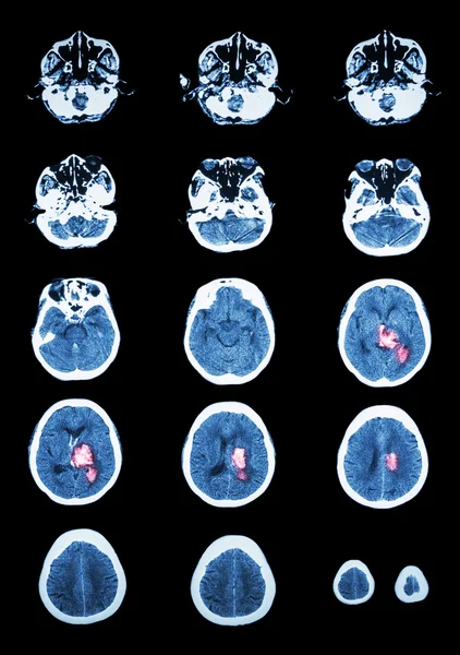
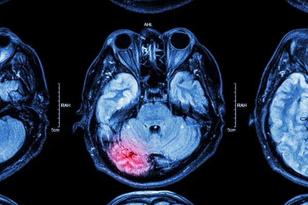
A CT Brain Scan Reveals A Subarachnoid Hemorrhage With Clear Signs Of Bleeding.
Image, 1.8MB, 6096 × 3215 jpg

A CT Brain Scan Reveals A Subarachnoid Hemorrhage With Clear Signs Of Bleeding.
Image, 3.06MB, 3244 × 4080 jpg

Kyiv, Ukraine - August 06, 2024: Studio Shoot Of Ferrer Takeda Citicoline Ceraxon Solution For Injections Ampoules Package Closeup On Black.
Image, 9.06MB, 5119 × 3412 jpg

Xray Of Head And Neck Isolated On Blue Backdround. The Joints And Bones,human Joints, Skeletal Spinal Bone Structure Of Human Spine, Medical Health Care Flat Vector Illustration.
Vector, 5.75MB, 5278 × 5278 eps

Selective Focus Of MRI Brain Sagittal Plane For Detect A Variety Of Conditions Of The Brain Such As Tumors. Idea Concept.
Image, 7.63MB, 6960 × 5256 jpg


Concept Photo Of Neurology And Diagnosis Of Cerebrovascular Diseases Such As Stroke, Encephalopathy, Cerebral Circulatory Insufficiency. Doctor In White Coat Holding Neurological Hammer, Stethoscope
Image, 5.82MB, 6000 × 4000 jpg
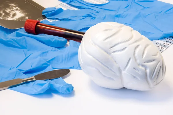
Brain And Nervous Surgery Or Neurosurgery Concept. Model Of Brain Near Scalpel, Ultrasound Result, Surgical Gloves And Blood Test Tube With Blood Result. Indications Neurosurgery Or Surgical Operation
Image, 7.14MB, 6000 × 4000 jpg


Film MRI Of Brain With Brain Tumor ( Sagittal Plane , Side View , Lateral View ) ( Medical , Health Care , Science Background )
Image, 3.36MB, 3096 × 3096 jpg

Stroke ( Cerebrovascular Accident ) . Film X-ray Skull Of Human With Red Area . Front View
Image, 3.49MB, 4779 × 4779 jpg


Cerebral Angiography Image From Fluoroscopy In Intervention Radiology Showing Cerebral Artery.
Image, 6.23MB, 4731 × 3845 jpg
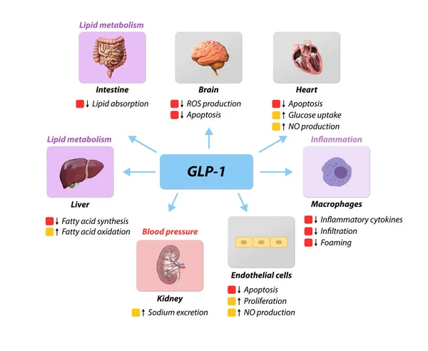
Glucagon-like Peptide 1 GLP-1 Prevents Macrovascular Complications,coronary Artery Disease, , Lipid Metabolism, Blood Pressure Inflammation, Nitric Oxide ROS, Oxygenmolecule, 2d 3d Graphic Rendering
Image, 2.47MB, 5928 × 4860 jpg
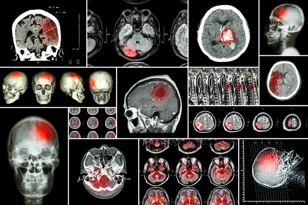
Set , Collection Of Brain Disease ( Cerebral Infarction , Hemorrhagic Stroke , Brain Tumor , Disc Herniation With Spinal Cord Compression ,etc)( CT Scan , MRI , MRT )( Neurology And Nervous System )
Image, 9.9MB, 6000 × 4000 jpg

Conceptual Hand Writing Text Caption Inspiration Showing Stroke Business Concept For Medicine Health Stethoscope Illness Written On Sticky Note Sculpture Background With Space
Image, 2.8MB, 5616 × 3744 jpg
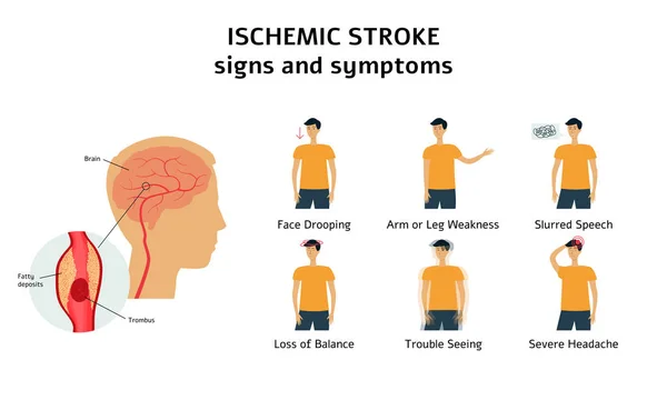
Ischemic Stroke Signs And Symptoms Infographic Flat Vector Illustration Isolated.
Vector, 6.12MB, 10417 × 6251 eps
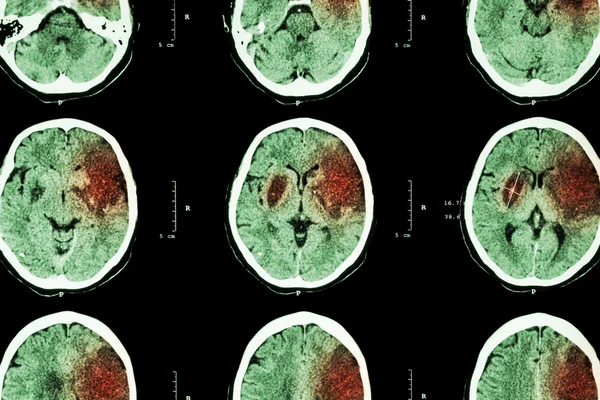
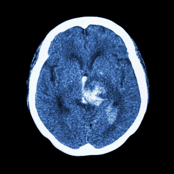
Ischemic Stroke : ( CT Of Brain Show Cerebral Infarction At Left Frontal - Temporal - Parietal Lobe ) ( Nervous System Background )
Image, 7.35MB, 5156 × 3437 jpg

Full Length Portrait Of Young Female Nurse Assisting Senior Patient With Remedial Bars. Old Man Doing Rehabilitation Between Parallel Bars.
Image, 11.26MB, 3648 × 5472 jpg

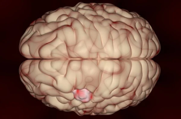
Meningioma (brain Cancer) Tumor In The Brain Tissue - 3d Illustration Top View
Image, 11.22MB, 10000 × 6600 jpg
Previous << Page 3 >> Next