
Stock image Ciliated

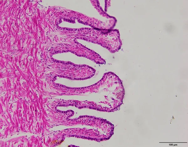
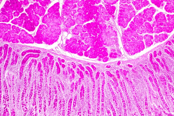
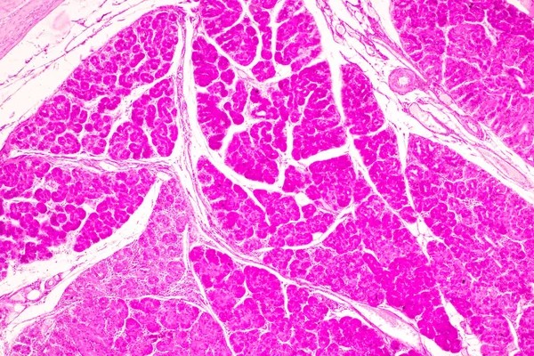
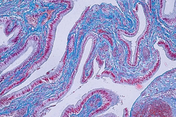
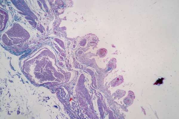
Tissue Of Small Intestine (Duodenum), Large Intestine Human And Stomach Human Under The Microscope In Lab.
Image, 17.29MB, 8192 × 5461 jpg
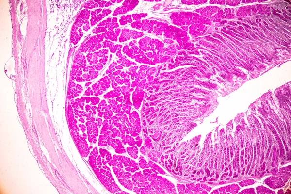
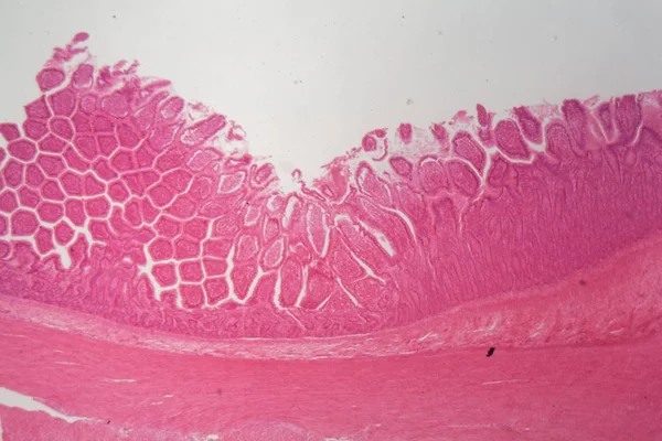
Tissue Of Small Intestine (Duodenum) And Vermiform Appendix Human Under The Microscope In Lab.
Image, 22.69MB, 6000 × 4000 jpg

Tissue Of Small Intestine (Duodenum) And Vermiform Appendix Human Under The Microscope In Lab.
Image, 20.31MB, 6000 × 4000 jpg

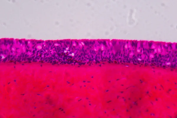
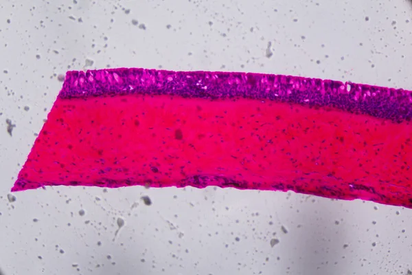
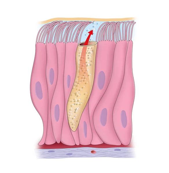
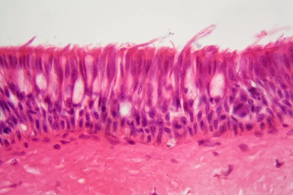
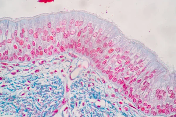
Ciliated Pseudostratified Columnar Epithelium Of The Trachea (respiratory Epithelium). The Apical Border Of The Epithelium Has A Layer Of Cilia (hair-like) Anchored In Their Basal Bodies. Among Ciliated Cells, Some Goblet Cells Can Be Seen.
Image, 8.55MB, 3840 × 3072 jpg

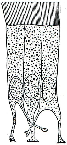
Phlox Drummondii Is A Flowering Plant. The Branches Have Sharp, Pointed, Lengthy, Ciliated Leaves With Rounded Flowers. The Flowers Are Single Or Double Star-shaped Petals, Vintage Line Drawing Or Engraving Illustration.
Vector, 5.06MB, 5016 × 6083 eps

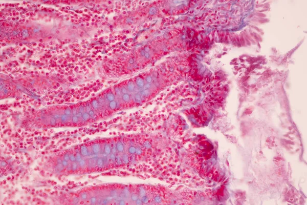
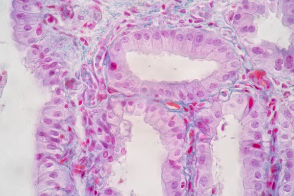
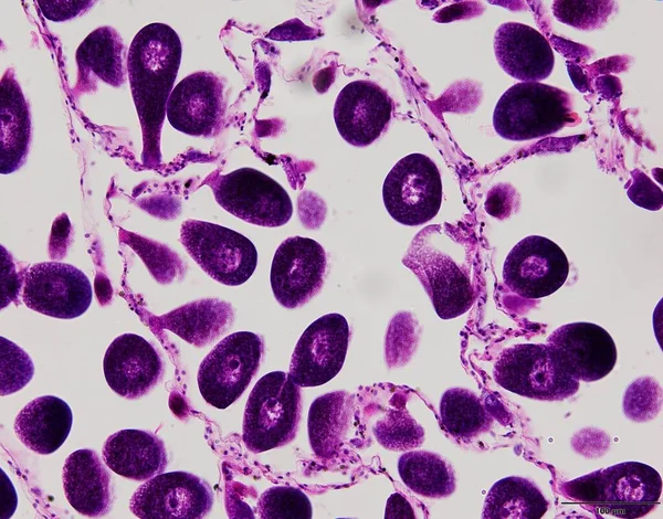
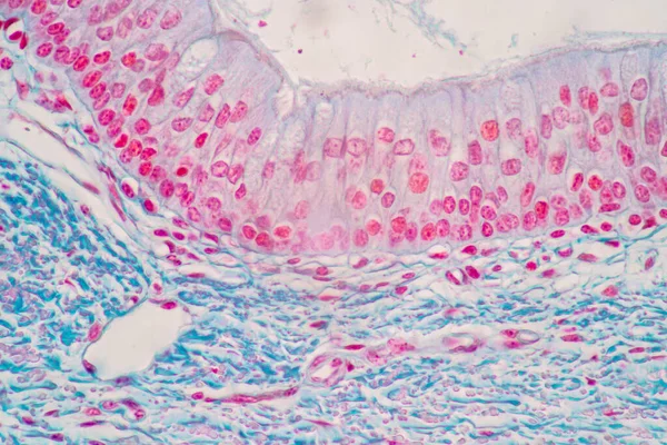
Characteristics Of Columnar Epithellum Cell (Cell Structure) Of Human Under Microscope View For Education In Laboratory.
Image, 16.38MB, 6720 × 4480 jpg
Tissue Of Small Intestine (Duodenum) And Vermiform Appendix Human Under The Microscope In Lab.
Image, 21.54MB, 6000 × 4000 jpg
Tissue Of Small Intestine (Duodenum) And Vermiform Appendix Human Under The Microscope In Lab.
Image, 18.99MB, 6000 × 4000 jpg

Characteristics Of Columnar Epithellum Cell (Cell Structure) Of Human Under Microscope View For Education In Laboratory.
Image, 17.03MB, 6720 × 4480 jpg
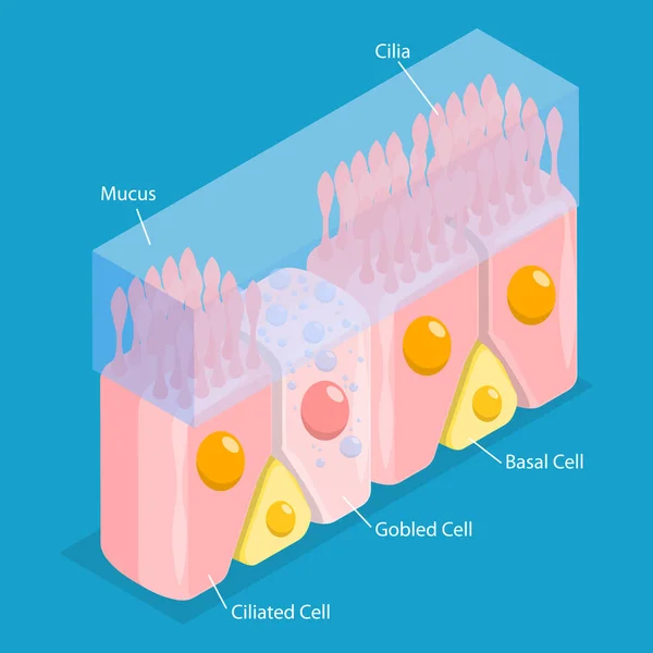
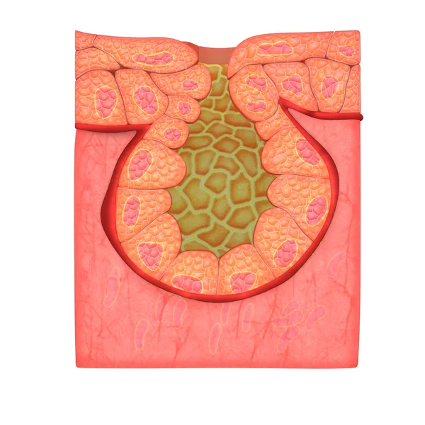
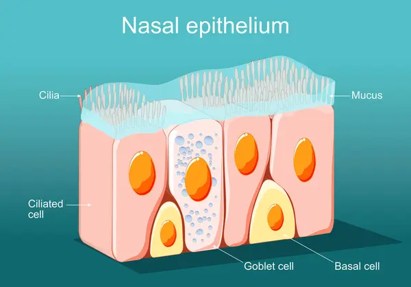
3D Isometric Flat Vector Conceptual Illustration Of Nasal Mucosa Cells, Medical Educational Diagram
Vector, 1.89MB, 5000 × 5000 eps

Ciliated Pseudostratified Columnar Epithelium Of The Trachea (respiratory Epithelium). The Apical Border Of The Epithelium Has A Layer Of Cilia (hair-like) Anchored In Their Basal Bodies. Among Ciliated Cells, Some Goblet Cells Can Be Seen.
Image, 9.74MB, 3840 × 3072 jpg


Characteristics Of Columnar Epithellum Cell (Cell Structure) Of Human Under Microscope View For Education In Laboratory.
Image, 16.28MB, 6720 × 4480 jpg
Characteristics Of Columnar Epithellum Cell (Cell Structure) Of Human Under Microscope View For Education In Laboratory.
Image, 17.31MB, 6720 × 4480 jpg


Tissue Of Small Intestine (Duodenum) And Vermiform Appendix Human Under The Microscope In Lab.
Image, 17.16MB, 6000 × 4000 jpg
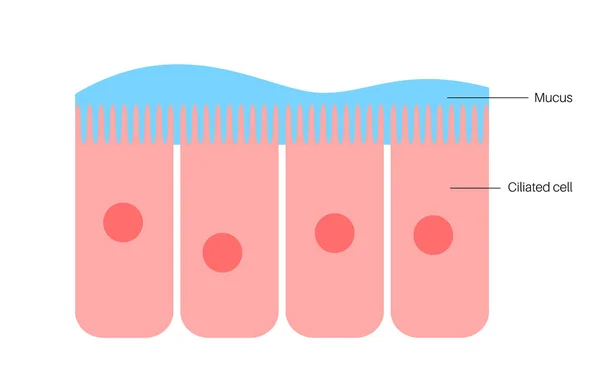

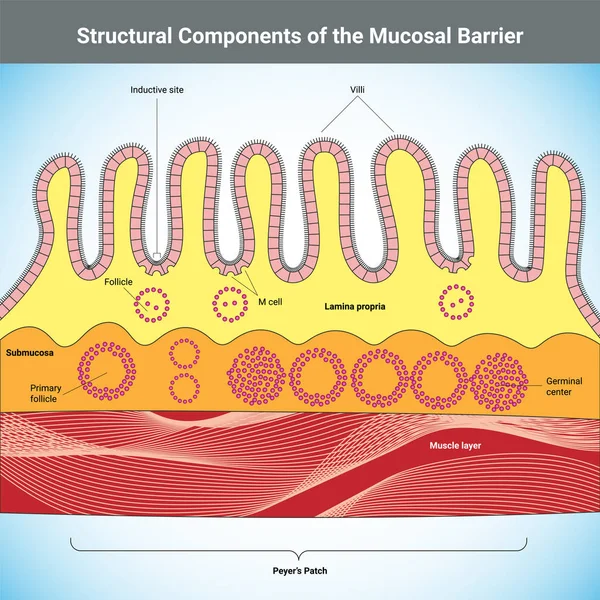
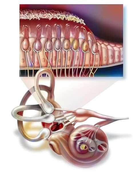
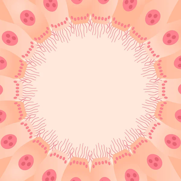
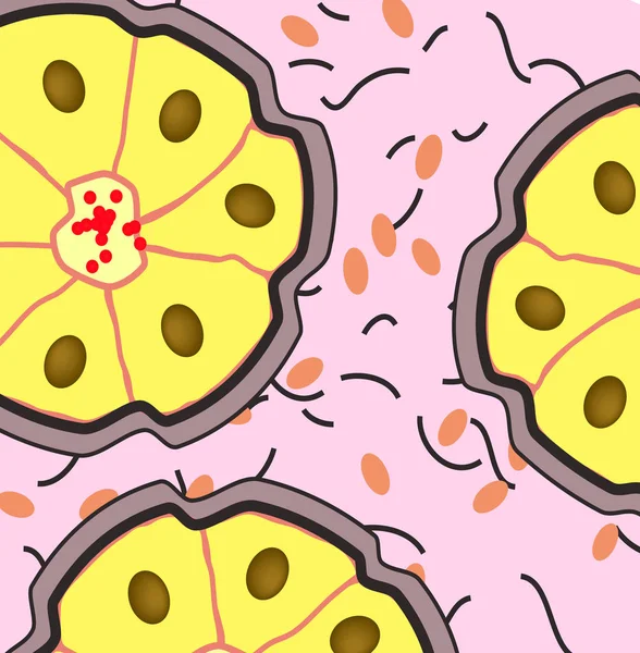
Nasal Epithelium. Ciliated Columnar Epithelium. Epithelial Cells Forms The Lining Of The Stomach And Intestines, Duodenum, Fallopian Tubes, Uterus, Central Canal Of The Spinal Cord, Nose, Ears And The Taste Buds. Ciliated Cells. Respiratory Defense
Vector, 2.23MB, 5000 × 3498 eps
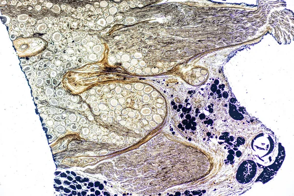
Showing Light Micrograph Type Of Tissue Human Under The Microscope In Lab.
Image, 25.49MB, 6000 × 4000 jpg
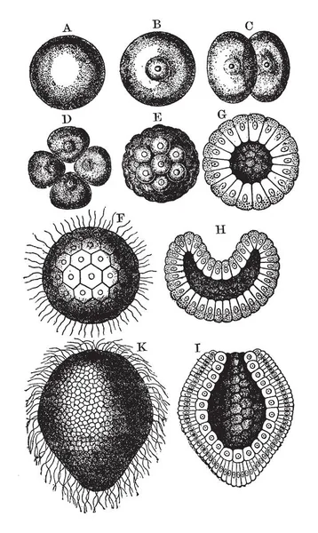
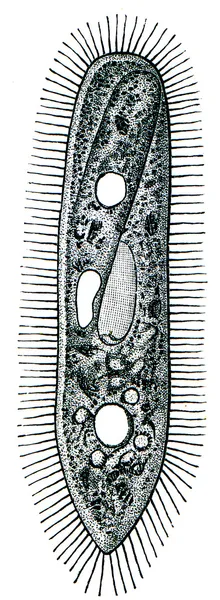
Coral Stages Where Free Swimming Ciliiated Gastrulais Another Labels, Vintage Line Drawing Or Engraving Illustration.
Vector, 8.54MB, 6100 × 10206 eps

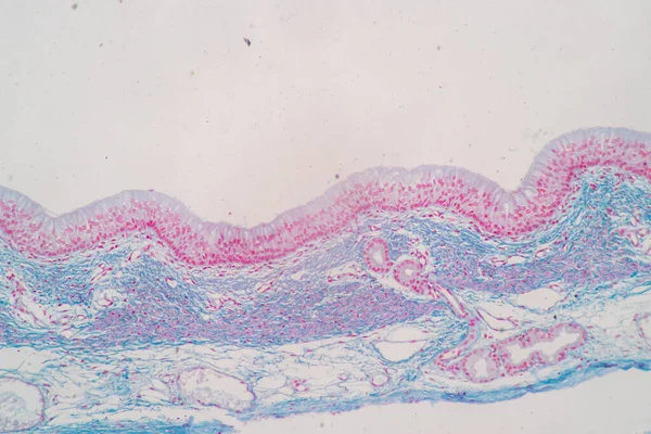
Cross Section Of Ciliated Epithelium Under The Microscope For Education Histology. Human Tissue.
Image, 14.75MB, 5168 × 3448 jpg
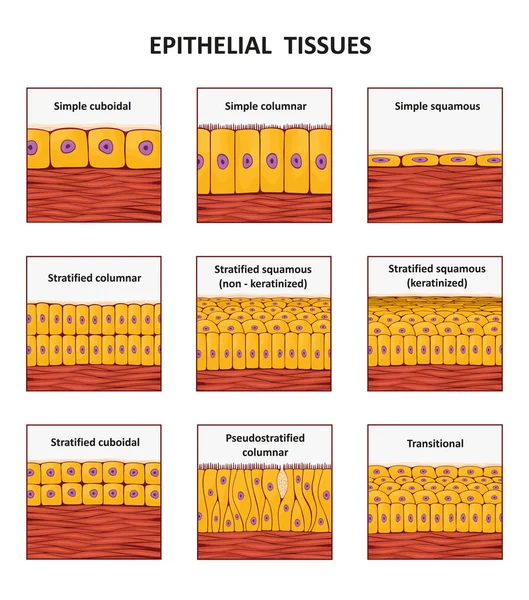
Epithelium. Squamous, Cubic, Ciliated, Glandular. Set. Infographics. Vector Illustration.
Vector, 1.56MB, 5000 × 5093 eps
Characteristics Of Columnar Epithellum Cell (Cell Structure) Of Human Under Microscope View For Education In Laboratory.
Image, 18.67MB, 6720 × 4480 jpg
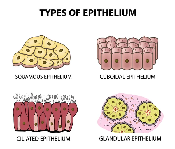
Types Of Epithelium. Squamous, Cubic, Ciliated, Glandular. Set. Infographics. Vector Illustration On Isolated Background
Vector, 2.27MB, 5000 × 4253 eps

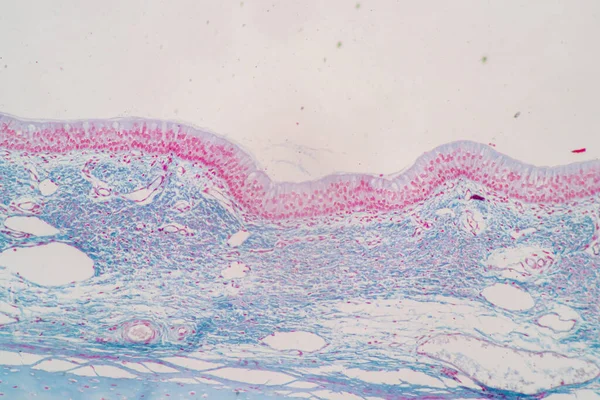
Pseudostratified Epithelium Is A Type Of Epithelium That, Though Comprising Only A Single Layer Of Cells.
Image, 11.24MB, 5840 × 3893 jpg


Characteristics Of Columnar Epithellum Cell (Cell Structure) Of Human Under Microscope View For Education In Laboratory.
Image, 16.3MB, 6720 × 4480 jpg
Characteristics Of Columnar Epithellum Cell (Cell Structure) Of Human Under Microscope View For Education In Laboratory.
Image, 17.33MB, 6720 × 4480 jpg
Page 1 >> Next