Home
Images
Vectors
Videos
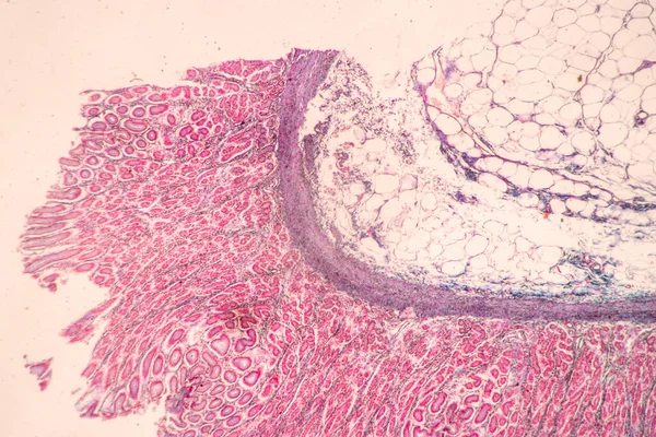
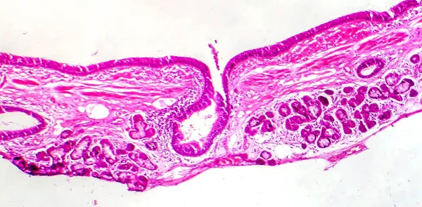
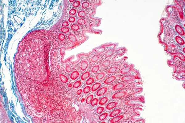
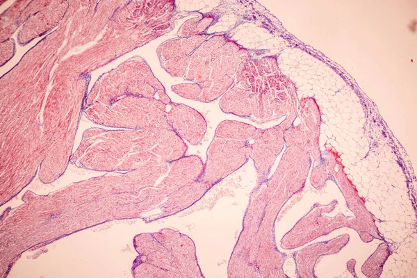
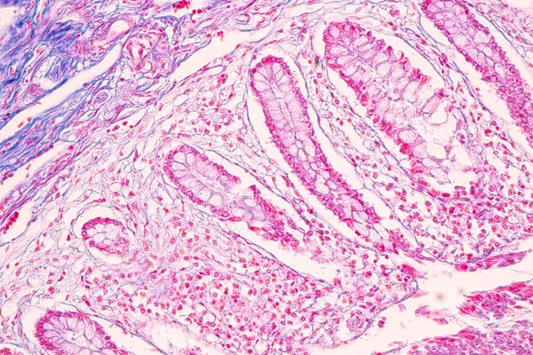
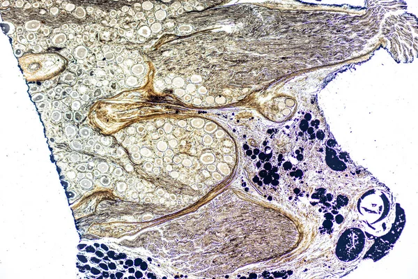
Stock image Showing Light micrograph Type of Tissue Human under the microscope in Lab.
Published:
Oct.20, 2023 12:51:29
Author:
p.thongdumhyu
Views:
3
Downloads:
1
File type:
image
/
jpg
File size:
25.49 MB
Orginal size:
6000
x
4000
px
Available sizes:
S
(500 x 333)
M
(1000 x 667)
L
(2000 x 1333)
XL
(6000 x 4000)
EL0
(6000 x 4000)
Level:
beginner
Download Image
Image topics:
Background
Human
Pattern
Smooth
Structure
Cell
Bone
Simple
Sample
Stained
Biology
Science
Fiber
Tissue
Striated
Muscle
Nucleus
Anatomy
Digestive
Microscope
Hyaline
Nervous
Absorption
Elastic
Microscopic
Skeletal
Stratified
Pathology
Nerve
Cilia
Microscopy
Columnar
Villi
Duodenum
Adipose
Intestinal
Cartilage
Pancreatic
Pathologist
Histology
Squamous
Epithelium
Microvilli
Squamous Cell
Connective Tissue
Cell Structure
Epithelial Cells
Ciliated
Columnar Epithelium
Simple Columnar Epithelium
Similar stock images
Backgrounds Of Characteristics Tissue Of Stomach Human, Small Intestine Human, Pancreas Human And Large Intestine Human Under The Microscope In Lab.
6720 × 4480
Showing Light Micrograph Type Of Tissue Human Under The Microscope In Lab.
6000 × 4000
Human Large Intestine Tissue Under Microscope View. Histological For Human Physiology.
6000 × 4000
Photomicrograph Of Bronchial Epithelial Squamous Metaplasia, Showing Squamous Cell Transformation In The Respiratory Lining.
4199 × 2071
Human Large Intestine Tissue Under Microscope View. Histological For Human Physiology.
6000 × 4000
Tissue Of Blood Human, Vein Human, Artery Human And Heart Muscle Human Under The Microscope In Lab.
8192 × 5464
Tissue Of Small Intestine (Duodenum) And Vermiform Appendix Human Under The Microscope In Lab.
6000 × 4000