
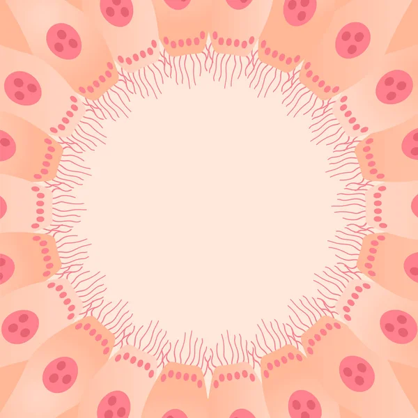
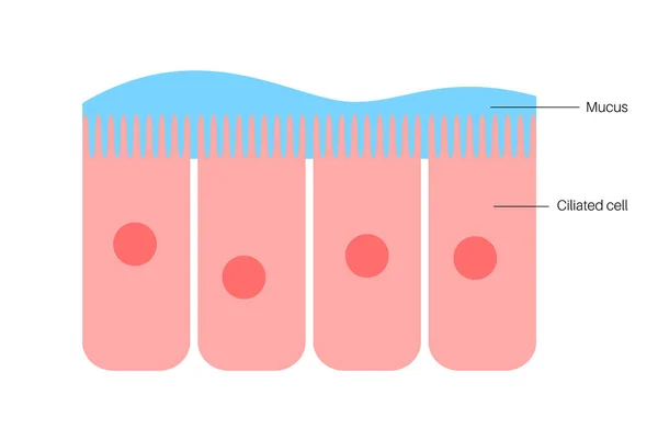
Stock image Ciliated Epithelium

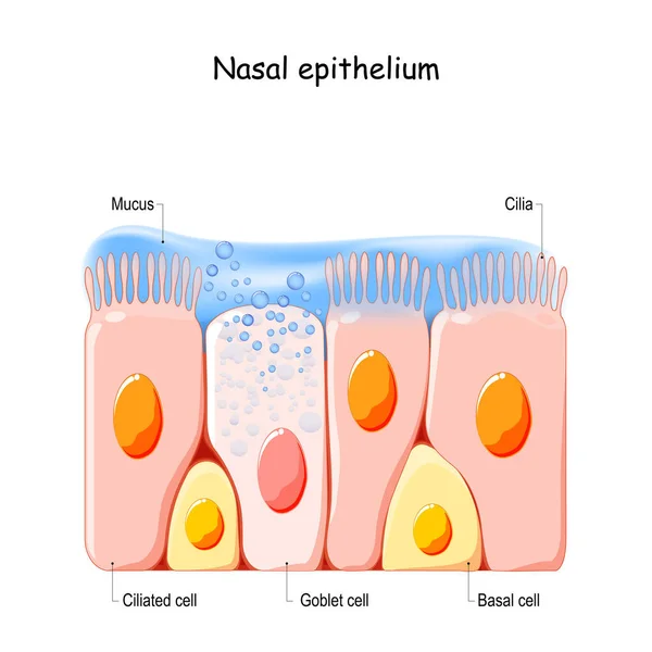
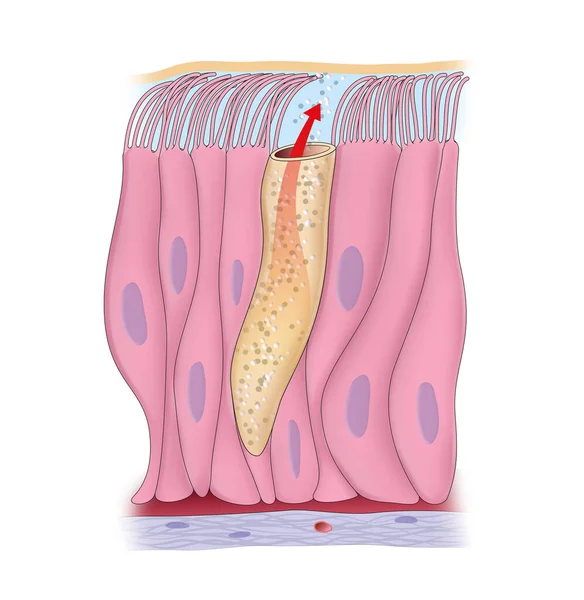
Nasal Mucosa Cells. Nasal Secretions. Ciliated, Basal And Goblet Cells. Olfactory Epithelium. Cells Act As A Low Resistance Filter. Vector Illustration
Vector, 11.58MB, 4444 × 4444 eps
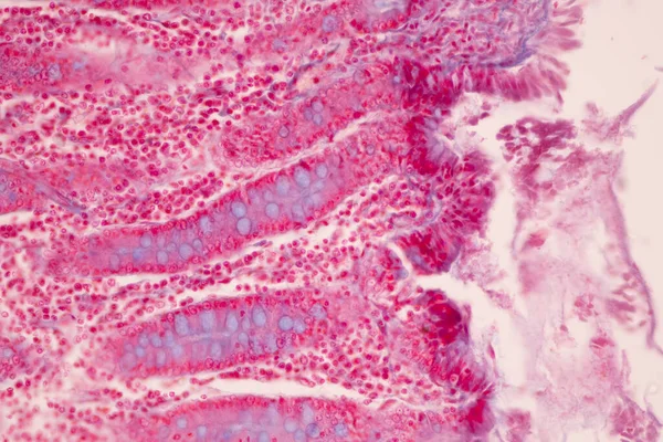
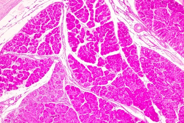
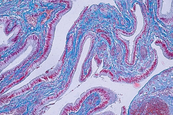
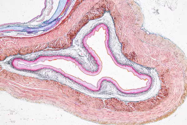
Tissue Of Small Intestine (Duodenum), Large Intestine Human And Stomach Human Under The Microscope In Lab.
Image, 17.29MB, 8192 × 5461 jpg
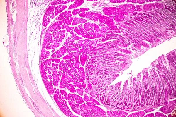
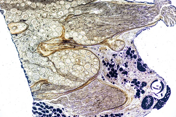
Tissue Of Small Intestine (Duodenum) And Vermiform Appendix Human Under The Microscope In Lab.
Image, 22.69MB, 6000 × 4000 jpg

Tissue Of Small Intestine (Duodenum) And Vermiform Appendix Human Under The Microscope In Lab.
Image, 20.31MB, 6000 × 4000 jpg
Tissue Of Small Intestine (Duodenum) And Vermiform Appendix Human Under The Microscope In Lab.
Image, 21.54MB, 6000 × 4000 jpg
Tissue Of Small Intestine (Duodenum) And Vermiform Appendix Human Under The Microscope In Lab.
Image, 18.99MB, 6000 × 4000 jpg


Tissue Of Small Intestine (Duodenum) And Vermiform Appendix Human Under The Microscope In Lab.
Image, 17.16MB, 6000 × 4000 jpg
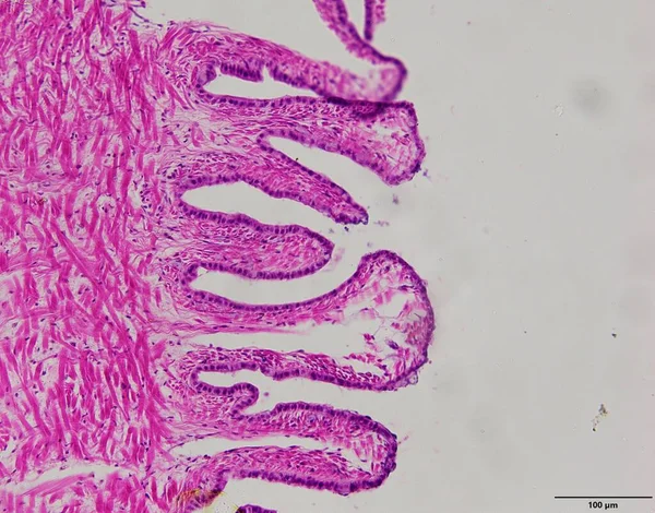
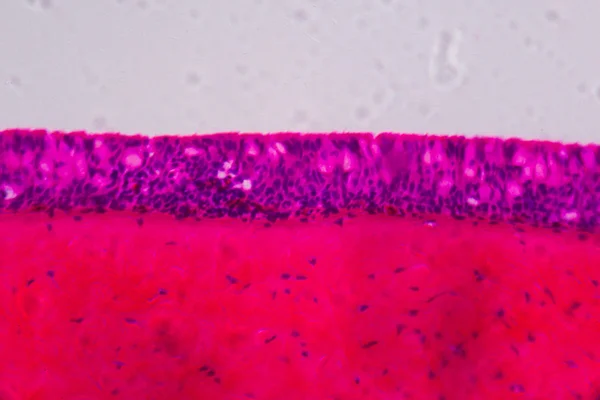
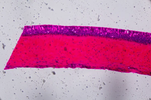
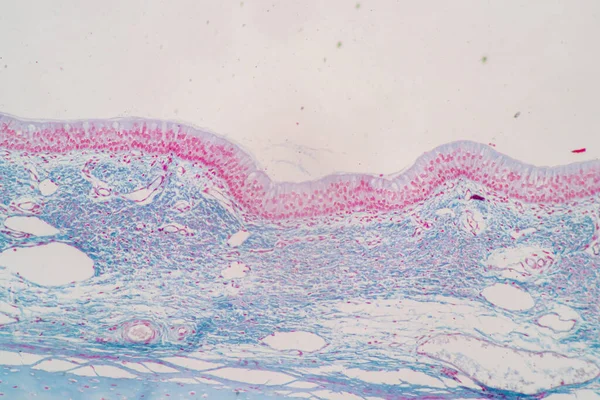
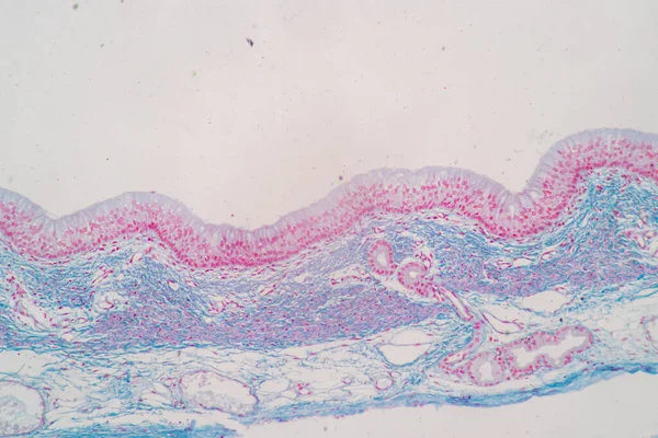
Cross Section Of Ciliated Epithelium Under The Microscope For Education Histology. Human Tissue.
Image, 14.75MB, 5168 × 3448 jpg

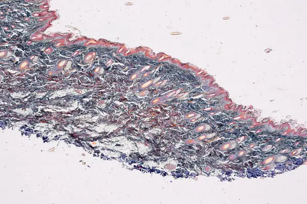
Pseudostratified Epithelium Is A Type Of Epithelium That, Though Comprising Only A Single Layer Of Cells.
Image, 11.24MB, 5840 × 3893 jpg


Ciliated Columnar Epithelium. Epithelial Cells Forms The Lining Of The Stomach And Intestines, Duodenum, Fallopian Tubes, Uterus, Central Canal Of The Spinal Cord, Nose, Ears And The Taste Buds.
Vector, 0.98MB, 4444 × 4444 eps


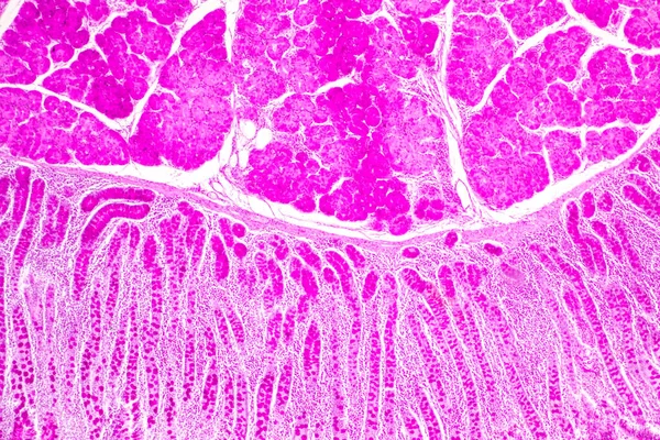
Pathology And Histology Tissue Of Mouse, Rabbit, Cat And Cow Under Microscope.
Image, 32.75MB, 6000 × 4000 jpg

Pathology And Histology Tissue Of Mouse, Rabbit, Cat And Cow Under Microscope.
Image, 6.85MB, 2667 × 4000 jpg
Pathology And Histology Tissue Of Mouse, Rabbit, Cat And Cow Under Microscope.
Image, 18.37MB, 6000 × 4000 jpg

Pathology And Histology Tissue Of Mouse, Rabbit, Cat And Cow Under Microscope.
Image, 8.03MB, 6000 × 3245 jpg


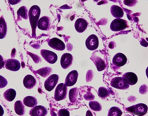
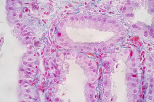
Characteristics Of Columnar Epithellum Cell (Cell Structure) Of Human Under Microscope View For Education In Laboratory.
Image, 16.38MB, 6720 × 4480 jpg

Pathology And Histology Tissue Of Mouse, Rabbit, Cat And Cow Under Microscope.
Image, 34.2MB, 6000 × 4000 jpg

Pathology And Histology Tissue Of Mouse, Rabbit, Cat And Cow Under Microscope.
Image, 18.99MB, 6000 × 4000 jpg
Characteristics Of Columnar Epithellum Cell (Cell Structure) Of Human Under Microscope View For Education In Laboratory.
Image, 17.03MB, 6720 × 4480 jpg
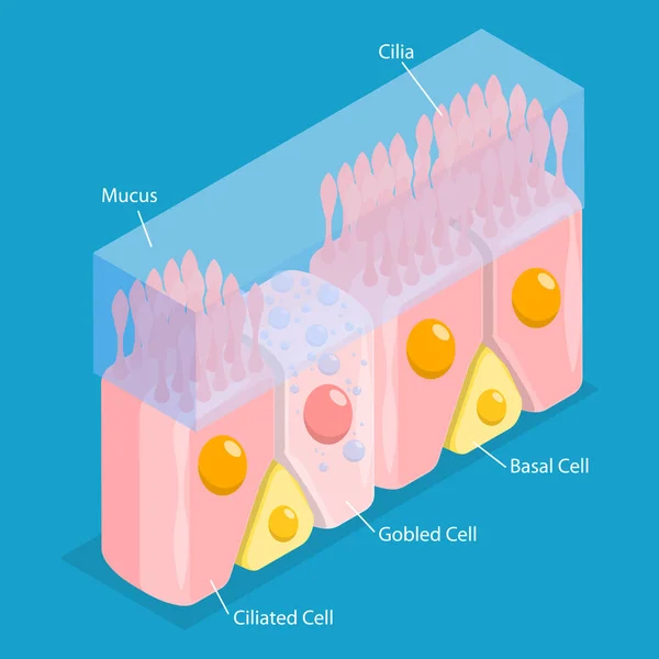
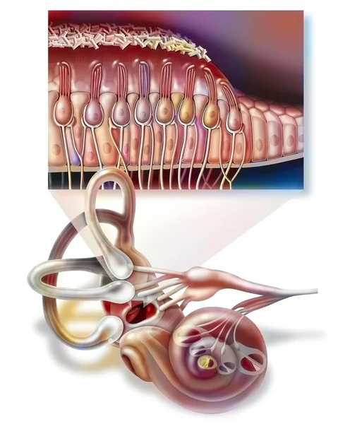
3D Isometric Flat Vector Conceptual Illustration Of Nasal Mucosa Cells, Medical Educational Diagram
Vector, 1.89MB, 5000 × 5000 eps

Pathology And Histology Tissue Of Mouse, Rabbit, Cat And Cow Under Microscope.
Image, 34.94MB, 6000 × 4000 jpg

Pathology And Histology Tissue Of Mouse, Rabbit, Cat And Cow Under Microscope.
Image, 10.76MB, 6000 × 4000 jpg

Pathology And Histology Tissue Of Mouse, Rabbit, Cat And Cow Under Microscope.
Image, 16.84MB, 6000 × 4000 jpg

Characteristics Of Columnar Epithellum Cell (Cell Structure) Of Human Under Microscope View For Education In Laboratory.
Image, 17.31MB, 6720 × 4480 jpg
Characteristics Of Columnar Epithellum Cell (Cell Structure) Of Human Under Microscope View For Education In Laboratory.
Image, 16.28MB, 6720 × 4480 jpg

Pathology And Histology Tissue Of Mouse, Rabbit, Cat And Cow Under Microscope.
Image, 20.89MB, 6000 × 4000 jpg
Pathology And Histology Tissue Of Mouse, Rabbit, Cat And Cow Under Microscope.
Image, 18.02MB, 6000 × 4000 jpg


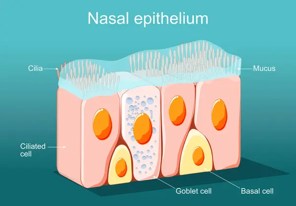
Nasal Epithelium. Ciliated Columnar Epithelium. Epithelial Cells Forms The Lining Of The Stomach And Intestines, Duodenum, Fallopian Tubes, Uterus, Central Canal Of The Spinal Cord, Nose, Ears And The Taste Buds. Ciliated Cells. Respiratory Defense
Vector, 2.23MB, 5000 × 3498 eps
Showing Light Micrograph Type Of Tissue Human Under The Microscope In Lab.
Image, 25.49MB, 6000 × 4000 jpg

Page 1 >> Next