
Stock image Clotting Cascade
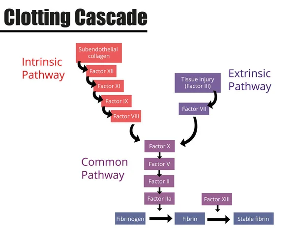
The Clotting Cascade Labeled Diagram. Coagulation Factors Of Blood. Vector Illustration. Didactic Illustration.
Vector, 0.65MB, 5000 × 4000 ai
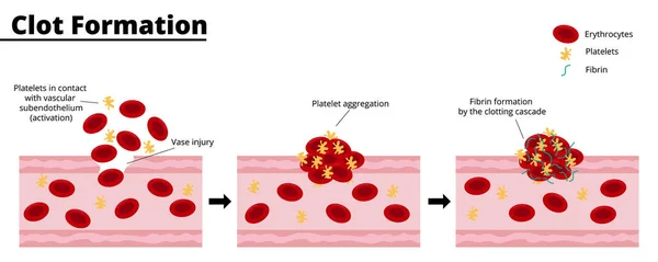
Clot Formation Process After A Bleeding Vascular Injury. Platelet Aggregate Formation. Formation Of Fibrin By The Clotting Cascade. Vector Illustration. Didactic Illustration.
Vector, 0.59MB, 7500 × 3000 ai
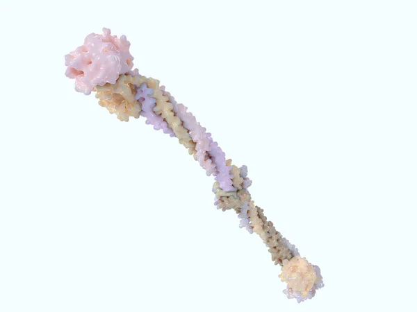
Fibrinogen Molecule. Fibrinogen Turns To Fibrin By The Catalysis Of Thrombin, Which Causes It To Polymerize. Fibrin Builds With Platelets A Clot Over Blood Vessel Injuries. 3D Rendering. Illustration
Image, 1.68MB, 8000 × 6000 jpg
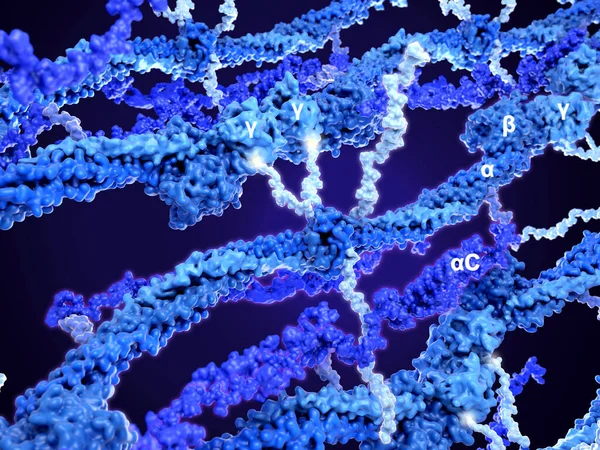
Structure Of A Fibrin Polymer. A Fibrin Monomer (highlighted) Is Composed Of 2, 2, 2 And 2C Domains. There Are 4 Flexible Chains (light Blue) In The Center. The 2 Short .chains Bind To Two Adjacent Domains Of The Polymer.
Image, 8.41MB, 8000 × 6000 jpg

Fibrinogen Molecule. Fibrinogen Turns To Fibrin By The Catalysis Of Thrombin, That Cleaves The Short Flexible Arms In The Middle Of The Molecule. Hence Fibrin Polymerizes Building Blood Clots. PDB Entries: 1m1j, 2baf.
Image, 4.23MB, 8000 × 6000 jpg

Heparin (yellow) Binds To And Activates Antithrombin. Antithrombin Inhibits Thrombin (dark Blue). The Anticoagulant Properties Of Heparin Are Mediated By Its Interaction With Antithrombin. Source: PDB Entry 1tb6
Image, 4.1MB, 8000 × 6000 jpg

2 Thrombin Molecules (red) Activate Fibrin By Cutting Some Aminoacid Residues (yellow Glow) From The Flexible Arms In The Middle Of The Fibrinogen Molecule. Hence Fibrin Polymerizes And Builds Blood Clots With Platelets. PDB Entries: 1m1j, 2baf, 2a45
Image, 4.63MB, 8000 × 6000 jpg

2 Thrombin Molecules (red) Activate Fibrin By Cutting Some Aminoacid Residues From The Flexible Arms In The Middle Of The Fibrinogen Molecule. Hence Fibrin Polymerizes And Builds Blood Clots With Platelets. PDB Entries: 1m1j, 2baf., 2a45
Image, 4.56MB, 8000 × 6000 jpg
Page 1 >> Next