
Stock image Descending Conduction

A 2:1 Left Bundle Branch Block Is Considered When Complete Left Bundle Branch Block Alternates With Normal QRS Complexes And The PR Interval Is Fixed.
Image, 5.72MB, 10000 × 3162 jpg
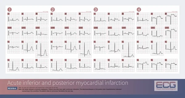
Male, 84 Years Old, Admitted To Hospital With Chest Pain For 1 Day. ECG Showed Acute Inferior And Posterior MI And Possibly Right MI. The Patient Died Of Ventricular Fibrillation The Next Day.
Image, 17.76MB, 10000 × 6934 jpg
Male, 84 Years Old, Admitted To Hospital With Chest Pain For 1 Day. These ECG Rhythms Are The Holter Monitor Records Of The Patients After Admission, And They Are Third Degree Atrioventricular Block.
Image, 16.1MB, 10000 × 5331 jpg

The Conduction In Ventricle Is Mainly Divided Into Right Bundle Branch And Left Bundle Branch. The Left Bundle Branch Includes Left Anterior Fascicle And Left Posterior Fascicle.
Image, 4.97MB, 10000 × 10000 jpg

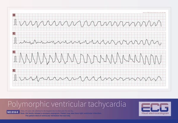
A Patient With AIMI Presents With A Sudden Widening Of The QRS Complex In The Junctional Escape Rhythm, Premature Ventricular Contractions, Resulting In Polymorphic Ventricular Tachycardia.
Image, 14.66MB, 10000 × 7554 jpg

The His Bundle And The Proximal Bundle Branches Are Mainly Supplied By The 1st Septal Branch Of The Left Anterior Descending Branch And The Atrioventricular Node Artery Of The Right Coronary Artery.
Image, 6.06MB, 10000 × 11566 jpg
Page 1 >> Next