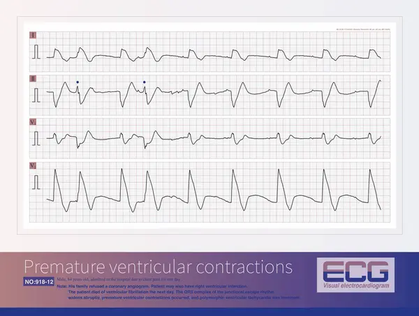
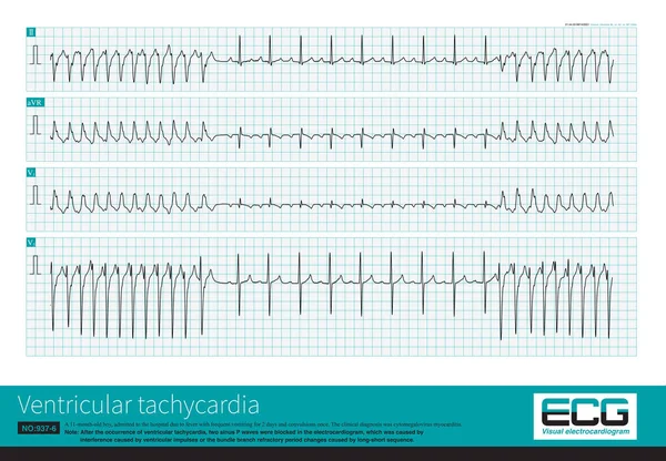
Stock image A patient with AIMI presents with a sudden widening of the QRS complex in the junctional escape rhythm, premature ventricular contractions, resulting in polymorphic ventricular tachycardia.

Published: Jul.20, 2024 13:34:04
Author: asia11m
Views: 0
Downloads: 0
File type: image / jpg
File size: 14.66 MB
Orginal size: 10000 x 7554 px
Available sizes:
Level: beginner