
Stock image Epithelial page 2

Mammary Cancer. Cancer Cells Under A Microscope. Tissues Affected By Cancer Cells Under A Microscope. Breast Cancer
Image, 5.39MB, 4184 × 2789 jpg
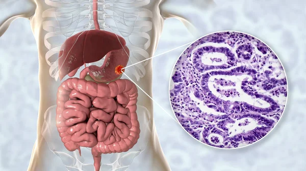
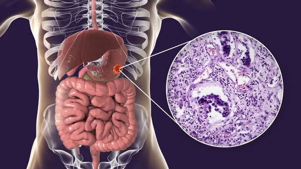
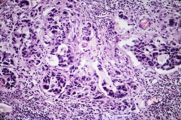
Stomach Adenocarcinoma, Gastric Cancer, Illustration And Light Micrograph
Image, 6.2MB, 5333 × 3000 jpg

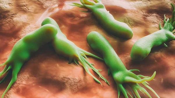
Cancer Cells Under A Microscope. Tissues Affected By Cancer Cells Under The Microscope, Science. Cancer Drugs. Chemistry And Biology.
Image, 7.04MB, 4854 × 2913 jpg

ACE - Angiotensin Converting Enzyme Acronym With Marker, Concept Background
Image, 3.22MB, 5760 × 3840 jpg
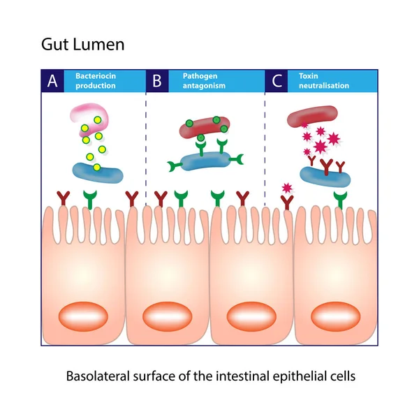
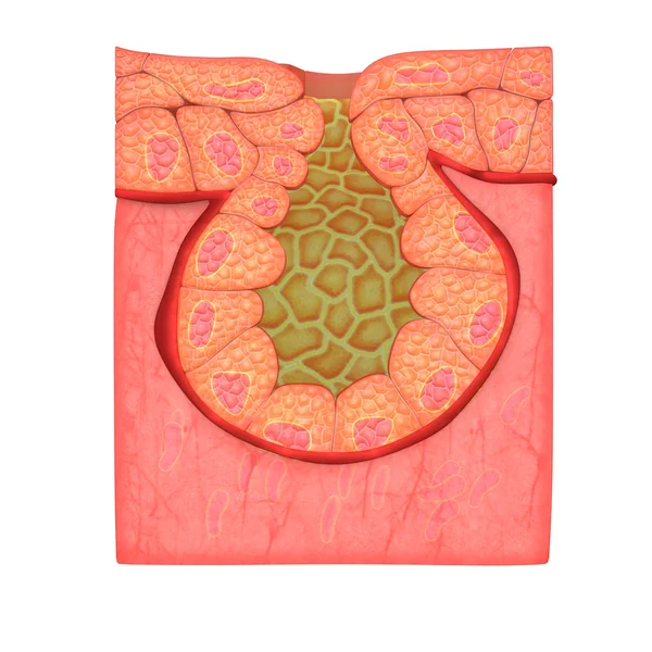
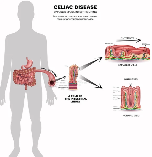
Gut Lumen. Enterocytes, Or Intestinal Absorptive Cells. Small Intestine. Columnar Epithelial Cells
Vector, 1.32MB, 5000 × 5000 eps

Squamous Epithelial Cells Under Microscope View For Education Histology. Human Tissue.
Image, 15.88MB, 6000 × 4000 jpg

Ovarian Cancer, 3D Illustration Showing Malignant Tumor In The Left Ovary
Image, 6.51MB, 6000 × 4000 jpg
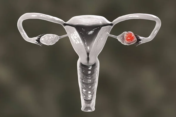
Ovarian Cancer, 3D Illustration Showing Malignant Tumor In The Left Ovary
Image, 4.45MB, 6000 × 4000 jpg
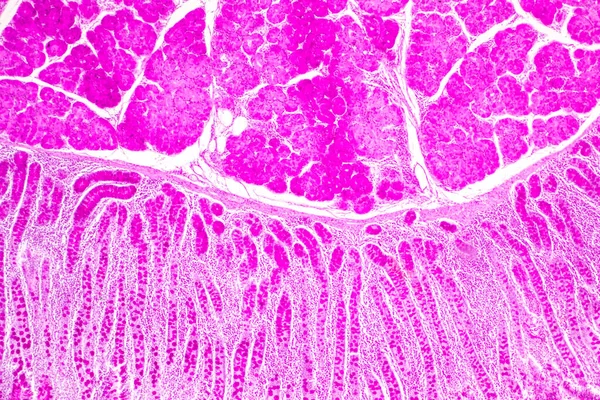
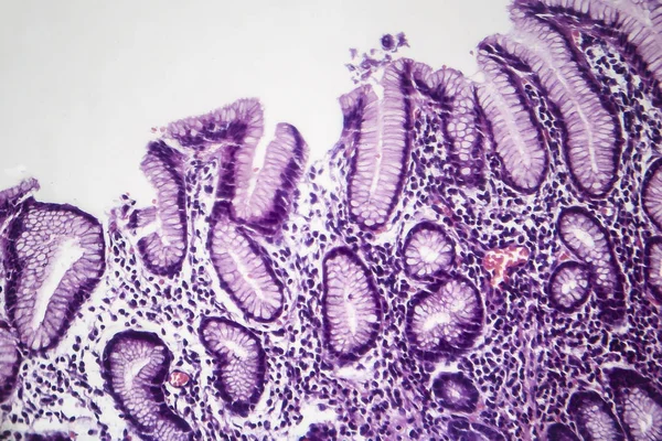
Tissue Of Small Intestine (Duodenum), Large Intestine Human And Stomach Human Under The Microscope In Lab.
Image, 17.29MB, 8192 × 5461 jpg
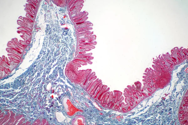
Tissue Of Small Intestine (Duodenum) And Vermiform Appendix Human Under The Microscope In Lab.
Image, 22.69MB, 6000 × 4000 jpg

Tissue Of Small Intestine (Duodenum) And Vermiform Appendix Human Under The Microscope In Lab.
Image, 20.31MB, 6000 × 4000 jpg


Stomach Adenocarcinoma, Gastric Cancer, Illustration And Light Micrograph
Image, 6.14MB, 5333 × 3000 jpg
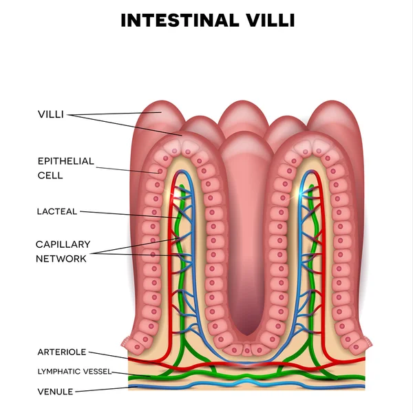
3d Illustration Intestinal Villi. Intestine Lining. Microscopic Villi And Capillary. Human Intestine. Viral Infection Causing Chronic Disease. Hepatitis Viruses, Influenza Virus, Cell Infect Organism.
Image, 6.7MB, 6000 × 4000 jpg

Intestinal Villi. Intestine Lining. Microscopic Capillary. Human Intestine. Concept Of A Healthy Or Diseased Intestinal. Viruses Or Bacteria, Cell Infected Organism, Decreased Immunity, 3D Rendering
Image, 12.45MB, 7500 × 5000 jpg
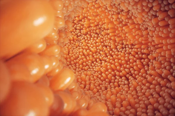
3D Illustration Close-up Intestinal Villi. Intestine Lining. Microscopic Villi And Capillary. Human Intestine. Concept Of A Healthy Or Diseased Intestine
Image, 15.32MB, 6000 × 4000 jpg

3D Illustration Close-up Intestinal Villi. Intestine Lining. Microscopic Villi And Capillary. Human Intestine. Concept Of A Healthy Or Diseased Intestine
Image, 11.39MB, 6000 × 4000 jpg
Human Cheek Epithelial Cells. The Tissue That Lines The Inside Of The Mouth Is Known As The Basal Mucosa And Is Composed Of Squamous Epithelial Cells. Education Pathology.
Image, 14.21MB, 6000 × 4000 jpg
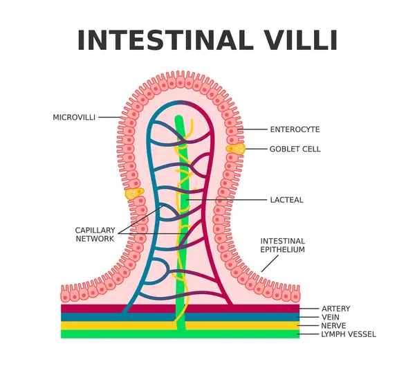
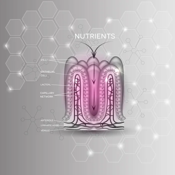
Intestinal Villi. Microvilli. Intestinal Epithelium. Villi Absorb Nutrients From The Food. Intestinal Epithelial Cells With Capillary Network. Enterocyte And Goblet Cell. Vector Illustration.
Vector, 9.49MB, 5000 × 4603 eps
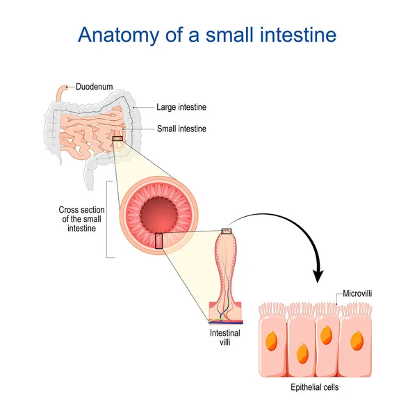
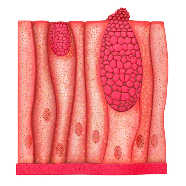
Small Intestine Anatomy. Cross Section Of A Ileum With Internal Villi. Close-up Of Epithelial Cells With Microvilli. Vector Illustration
Vector, 5.75MB, 4444 × 4444 eps
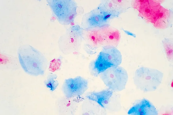
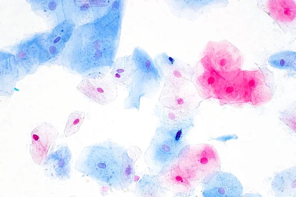
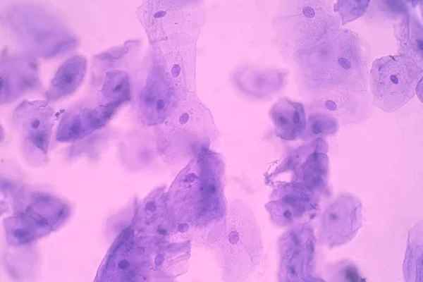
Squamous Epithelial Cells Of Human Cervix Under The Microscope View. Pap Smear Test Is A Procedure To Test For Cervical Cancer In Women.
Image, 17.23MB, 5883 × 3922 jpg

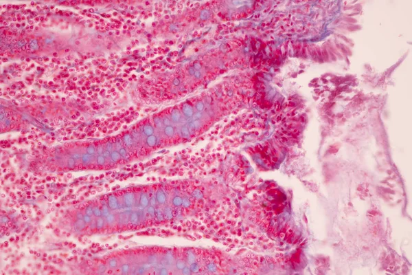
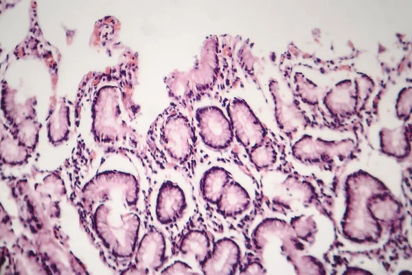
This Is A Histological Photograph Of The Human Small Intestine. Magnify 40x.
Image, 9.15MB, 4202 × 4202 jpg
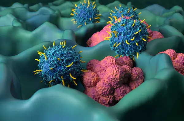
CAR T Cell Therapy In Lung Cancer (LC) - Closeup View 3d Illustration
Image, 8.33MB, 10000 × 6600 jpg


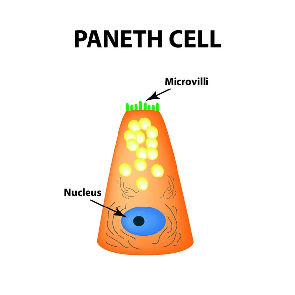
The Structure Of Paneth Cells. Davidoff's Cell. Fographics. Vector Illustration On Isolated Background.
Vector, 0.78MB, 5000 × 5000 eps
Human Large Intestine Tissue Under Microscope View. Histological For Human Physiology.
Image, 15.29MB, 6000 × 4000 jpg

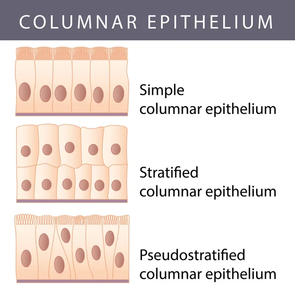
Characteristics Of Columnar Epithellum Cell (Cell Structure) Of Human Under Microscope View For Education In Laboratory.
Image, 16.38MB, 6720 × 4480 jpg

Stomach Adenocarcinoma, Gastric Cancer, Illustration And Light Micrograph
Image, 6.66MB, 5333 × 3000 jpg

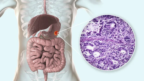
Stomach Adenocarcinoma, Gastric Cancer, Illustration And Light Micrograph
Image, 6.46MB, 5333 × 3000 jpg
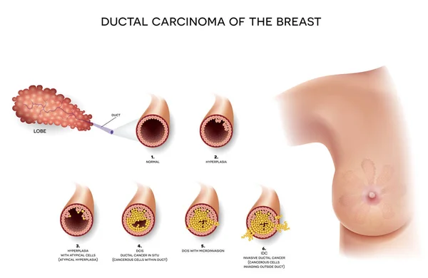
Ductal Carcinoma Of The Breast, Detailed Medical Illustration. At The Beginning Normal Duct, Then Hyperplasia, After That Atypical Cells Are Invading, Ductal Cancer In Situ And Invasive Ductal Cancer.
Vector, 10.79MB, 6050 × 3933 eps
Previous << Page 2 >> Next