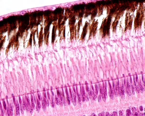
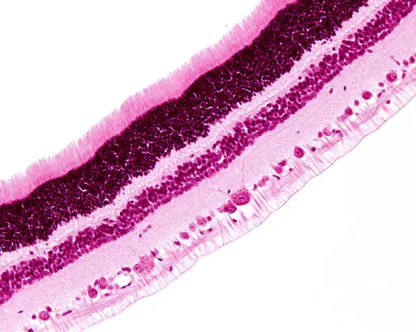
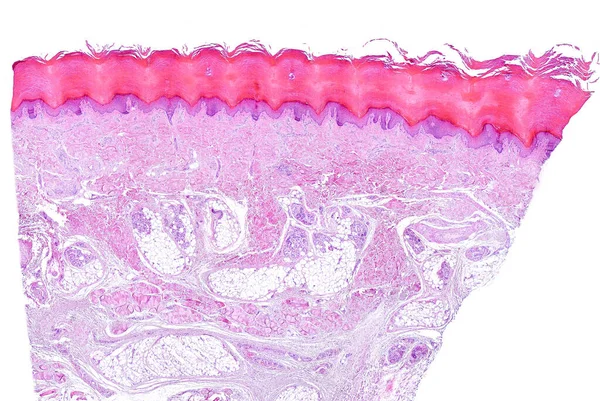
Stock image This is a histological photograph of the human small intestine. Magnify 40x.

Published: May.03, 2024 13:10:26
Author: asia11m
Views: 4
Downloads: 1
File type: image / jpg
File size: 9.15 MB
Orginal size: 4202 x 4202 px
Available sizes:
Level: beginner