
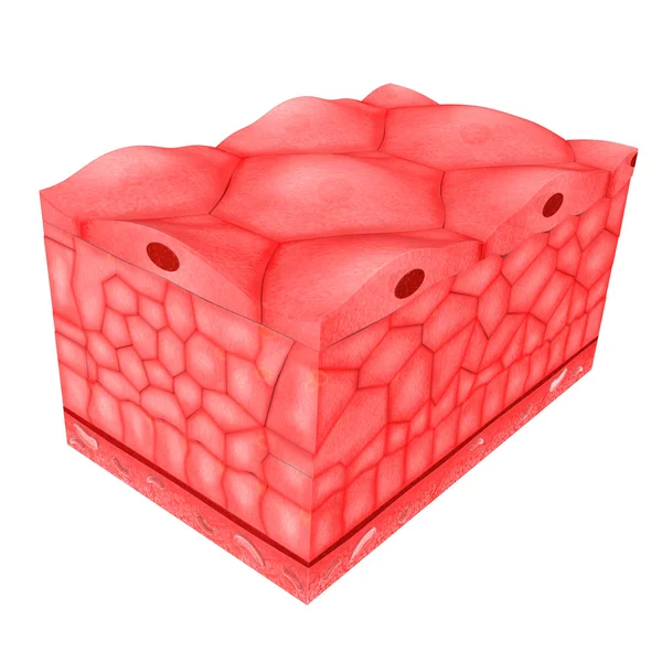
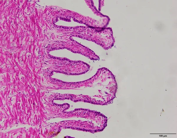
Stock image Epithelium

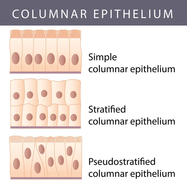

Ciliated Columnar Epithelium. Epithelial Cells Forms The Lining Of The Stomach And Intestines, Duodenum, Fallopian Tubes, Uterus, Central Canal Of The Spinal Cord, Nose, Ears And The Taste Buds.
Vector, 0.98MB, 4444 × 4444 eps
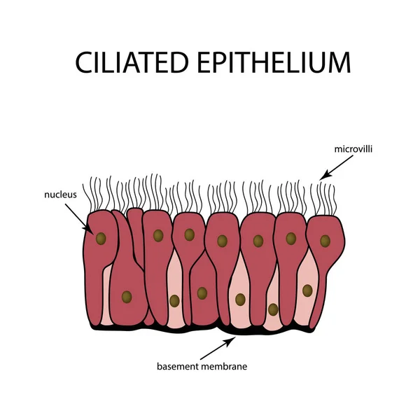
The Structure Of The Ciliated Epithelium. Infographics. Vector Illustration On Isolated Background
Vector, 0.49MB, 5000 × 5000 eps
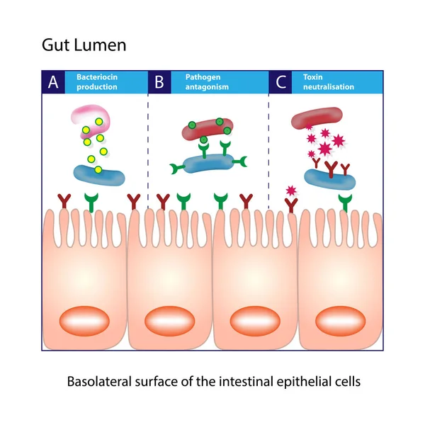
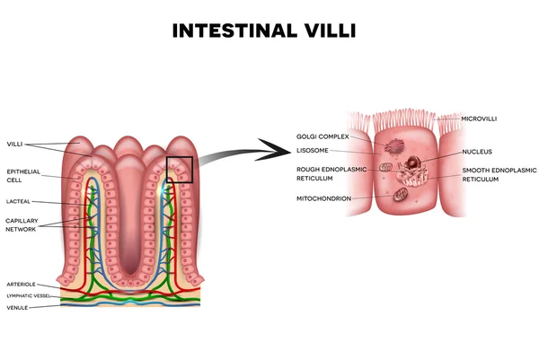
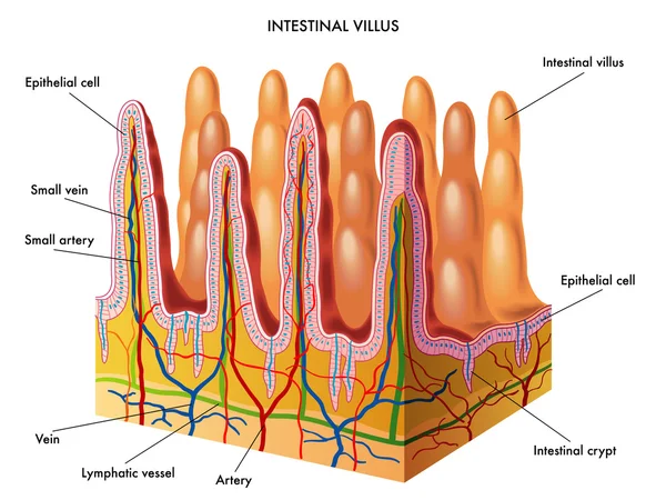
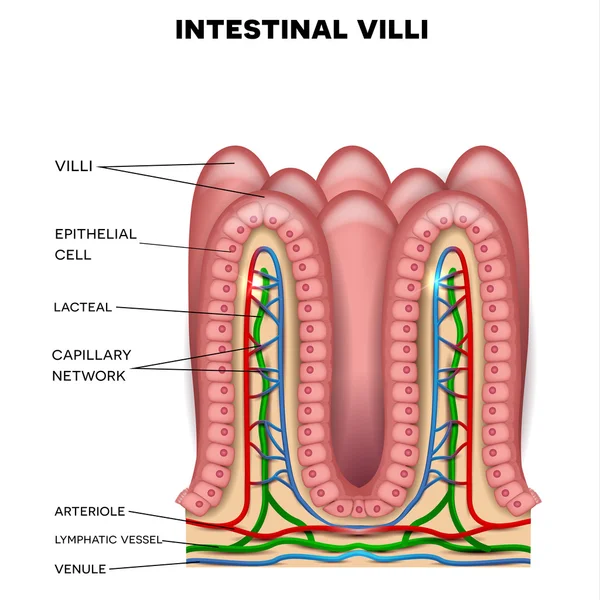
Gut Lumen. Enterocytes, Or Intestinal Absorptive Cells. Small Intestine. Columnar Epithelial Cells
Vector, 1.32MB, 5000 × 5000 eps

The Structure Of The Glandular Epithelium. Infographics. Vector Illustration On Isolated Background
Vector, 0.78MB, 5000 × 5000 eps
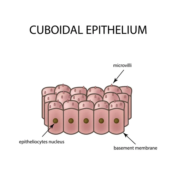
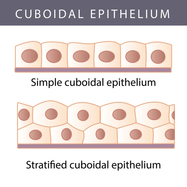
The Structure Of Cubic Epithelium. Infographics. Vector Illustration On Isolated Background
Vector, 1.62MB, 5000 × 5000 eps


25.07.2020, Zarorizhzhya. Topography Of The Cornea Of The Eye, Layout. Keratoconus And Glasses. Keratotopography - Ophthalmological Examination.
Image, 10.41MB, 6000 × 4000 jpg

Woman Holding Her Nose, Concept Anosmia Or Hyposma, Rhinaesthesia, Stench
Image, 6.11MB, 3516 × 5274 jpg


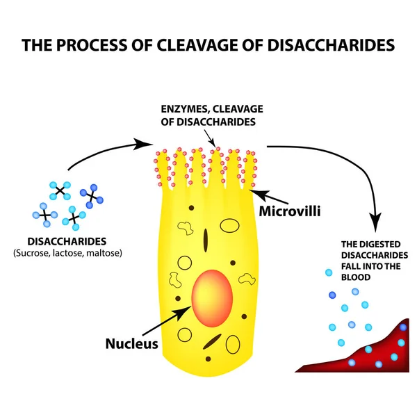
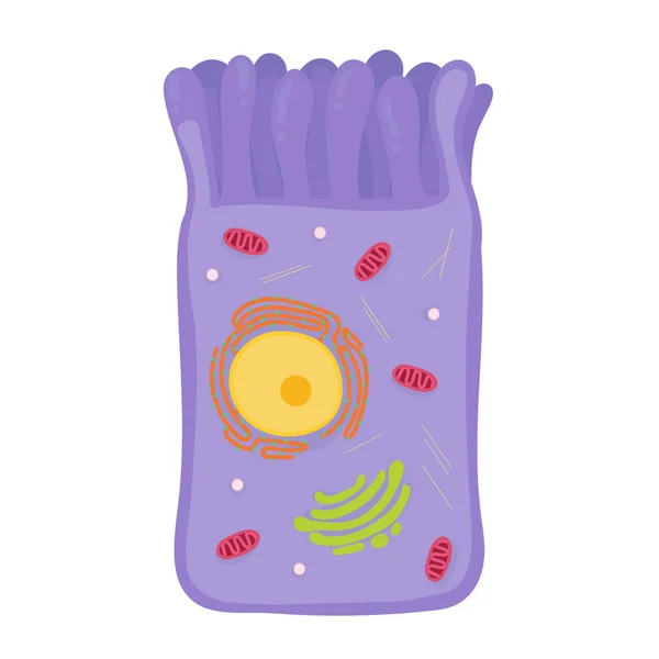
The Process Of Cleavage Of Disaccharides. Structure Of The Enterocyte. Absorptive Cells Intestine. Infographics. Vector Illustration On Isolated Background.
Vector, 2.09MB, 5000 × 5000 eps

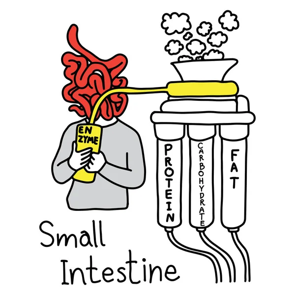
Metaphor Function Of Small Intestine To Make Enzyme To Digest Protein Fat And Carbohydrate Vector Illustration Sketch Hand Drawn With Black Lines, Isolated On White Background
Vector, 5.76MB, 5000 × 5000 eps

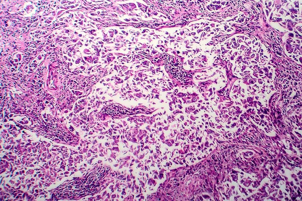
Uterus Adenofibroma, Light Micrograph, Photo Under Microscope. A Rare Benign Tumor Of The Uterus Composed Of Glandular And Fibrous Tissues
Image, 9.96MB, 4602 × 3068 jpg
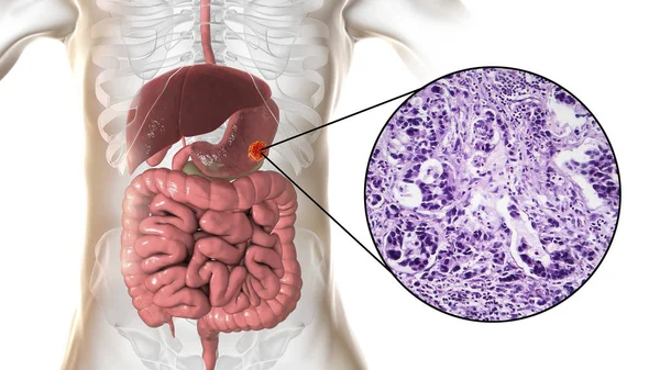
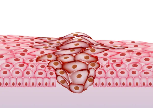
Stomach Adenocarcinoma, Gastric Cancer, Illustration And Light Micrograph
Image, 5.8MB, 5333 × 3000 jpg

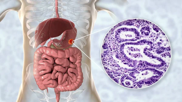
Stomach Adenocarcinoma, Gastric Cancer, Illustration And Light Micrograph
Image, 6.2MB, 5333 × 3000 jpg


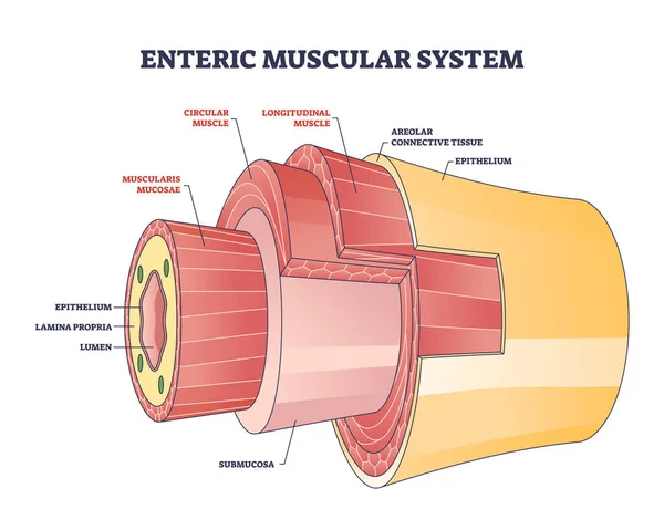
Enteric Muscular System In Gut Wall Of The Small Intestine Outline Diagram. Labeled Educational Scheme With Layers And Structure Of Digestive Tract Muscle Vector Illustration. Lamina Propria Location.
Vector, 7.06MB, 4500 × 3600 eps
Once An Atherosclerotic Plaque Reaches A Certain Size, It Can Occlude An Artery Completely Or Part Of The Plaque Can Rupture And Form A Clot, Causing Occlusion. 3D Rendering
Image, 19.22MB, 11115 × 5802 jpg


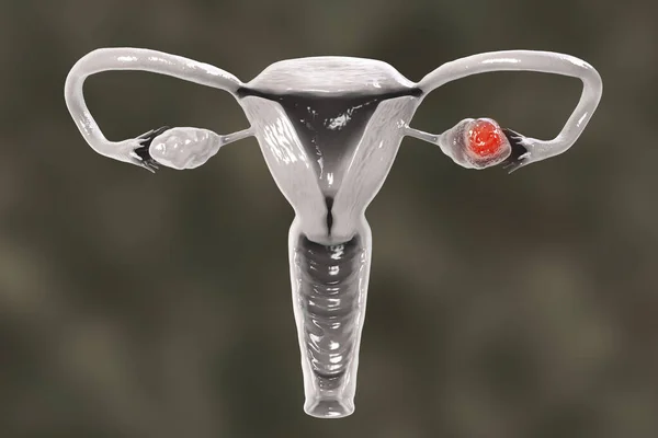
Ovarian Cancer, 3D Illustration Showing Malignant Tumor In The Left Ovary
Image, 6.51MB, 6000 × 4000 jpg
Ovarian Cancer, 3D Illustration Showing Malignant Tumor In The Left Ovary
Image, 4.45MB, 6000 × 4000 jpg
Page 1 >> Next