
Stock image Epithelium page 2
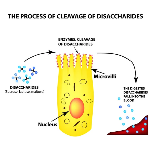
The Process Of Cleavage Of Disaccharides. Structure Of The Enterocyte. Absorptive Cells Intestine. Infographics. Vector Illustration On Isolated Background.
Vector, 2.09MB, 5000 × 5000 eps

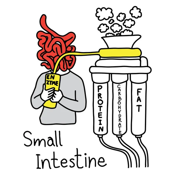
Metaphor Function Of Small Intestine To Make Enzyme To Digest Protein Fat And Carbohydrate Vector Illustration Sketch Hand Drawn With Black Lines, Isolated On White Background
Vector, 5.76MB, 5000 × 5000 eps

Uterus Adenofibroma, Light Micrograph, Photo Under Microscope. A Rare Benign Tumor Of The Uterus Composed Of Glandular And Fibrous Tissues
Image, 9.96MB, 4602 × 3068 jpg
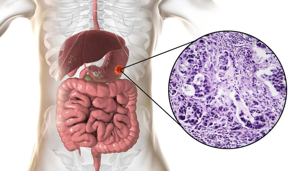
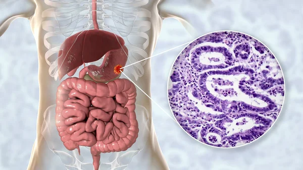
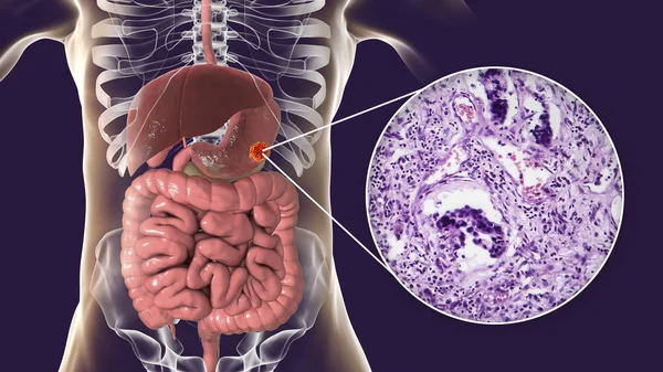
Stomach Adenocarcinoma, Gastric Cancer, Illustration And Light Micrograph
Image, 5.8MB, 5333 × 3000 jpg

Stomach Adenocarcinoma, Gastric Cancer, Illustration And Light Micrograph
Image, 6.2MB, 5333 × 3000 jpg


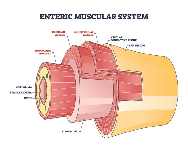
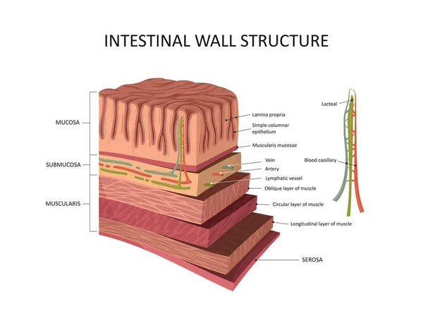
Enteric Muscular System In Gut Wall Of The Small Intestine Outline Diagram. Labeled Educational Scheme With Layers And Structure Of Digestive Tract Muscle Vector Illustration. Lamina Propria Location.
Vector, 7.06MB, 4500 × 3600 eps
Once An Atherosclerotic Plaque Reaches A Certain Size, It Can Occlude An Artery Completely Or Part Of The Plaque Can Rupture And Form A Clot, Causing Occlusion. 3D Rendering
Image, 19.22MB, 11115 × 5802 jpg


Ovarian Cancer, 3D Illustration Showing Malignant Tumor In The Left Ovary
Image, 6.51MB, 6000 × 4000 jpg
Ovarian Cancer, 3D Illustration Showing Malignant Tumor In The Left Ovary
Image, 4.45MB, 6000 × 4000 jpg
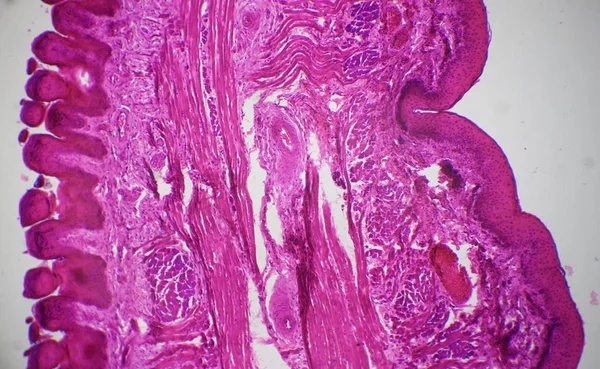
Tissue Of Small Intestine (Duodenum), Large Intestine Human And Stomach Human Under The Microscope In Lab.
Image, 17.29MB, 8192 × 5461 jpg


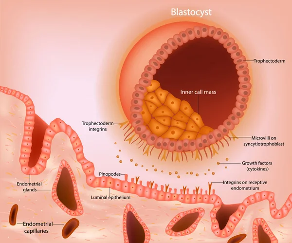
Blastocyst Implantation. A Schematic Representation Of A Blastocyst Approaching The Receptive Endometrium. Early Signaling Between The Blastocyst. Embryonic Development
Vector, 3.97MB, 6000 × 5000 eps
Stomach Adenocarcinoma, Gastric Cancer, Illustration And Light Micrograph
Image, 6.14MB, 5333 × 3000 jpg
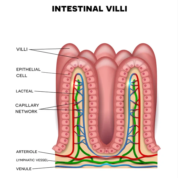
3d Illustration Intestinal Villi. Intestine Lining. Microscopic Villi And Capillary. Human Intestine. Viral Infection Causing Chronic Disease. Hepatitis Viruses, Influenza Virus, Cell Infect Organism.
Image, 6.7MB, 6000 × 4000 jpg

Intestinal Villi. Intestine Lining. Microscopic Capillary. Human Intestine. Concept Of A Healthy Or Diseased Intestinal. Viruses Or Bacteria, Cell Infected Organism, Decreased Immunity, 3D Rendering
Image, 12.45MB, 7500 × 5000 jpg
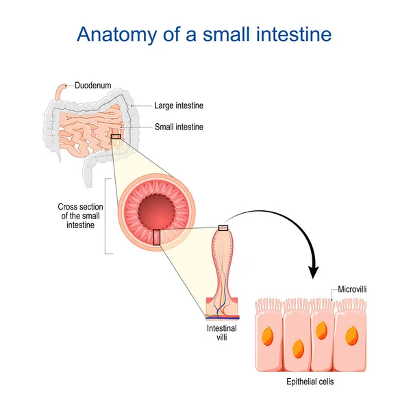
Small Intestine Anatomy. Cross Section Of A Ileum With Internal Villi. Close-up Of Epithelial Cells With Microvilli. Vector Illustration
Vector, 5.75MB, 4444 × 4444 eps

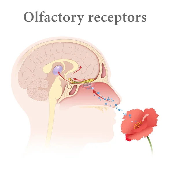
The Sense Of Smell Detects Airborne Molecules Via Olfactory Receptors In The Nasal Cavity, Sending Signals To The Brain For Perception
Image, 1.95MB, 5670 × 3971 jpg
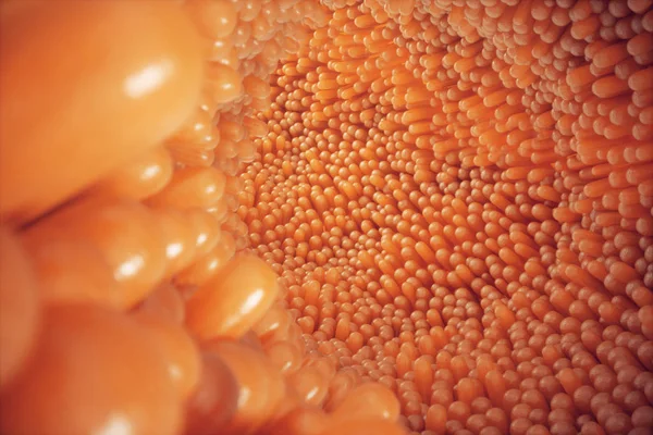
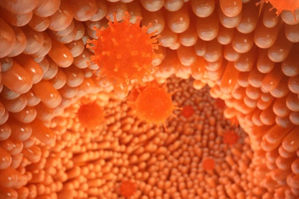
3D Illustration Close-up Intestinal Villi. Intestine Lining. Microscopic Villi And Capillary. Human Intestine. Concept Of A Healthy Or Diseased Intestine
Image, 15.32MB, 6000 × 4000 jpg


Cervical Cancer Cells Elongated Dysplasia Stages Infographic Female Reproductive System Squamous Cell Carcinoma In Uterus Pap Smear Test. Human Anatomy Internal Organs Flat Style Icon
Vector, 6.46MB, 10040 × 5002 eps
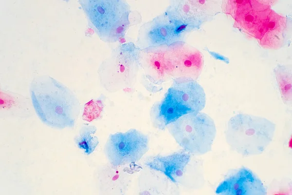
Squamous Epithelial Cells Of Human Cervix Under The Microscope View. Pap Smear Test Is A Procedure To Test For Cervical Cancer In Women.
Image, 17.23MB, 5883 × 3922 jpg
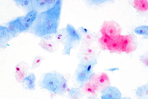
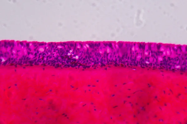
Squamous Epithelial Cells Under Microscope View For Education Histology. Human Tissue.
Image, 15.88MB, 6000 × 4000 jpg
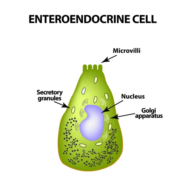
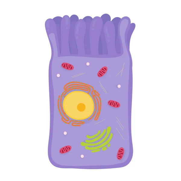
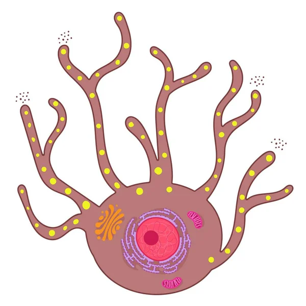
Enteroendocrine Cell. Cell Of The Intestines. Vector Illustration On Isolated Background
Vector, 0.97MB, 5000 × 5000 eps

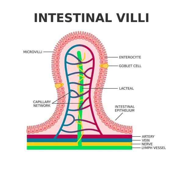
Intestinal Villi. Microvilli. Intestinal Epithelium. Villi Absorb Nutrients From The Food. Intestinal Epithelial Cells With Capillary Network. Enterocyte And Goblet Cell. Vector Illustration.
Vector, 9.49MB, 5000 × 4603 eps


3D Illustration Close-up Intestinal Villi. Intestine Lining. Microscopic Villi And Capillary. Human Intestine. Concept Of A Healthy Or Diseased Intestine
Image, 11.39MB, 6000 × 4000 jpg
Previous << Page 2 >> Next