
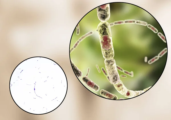
Stock image Gram Stain
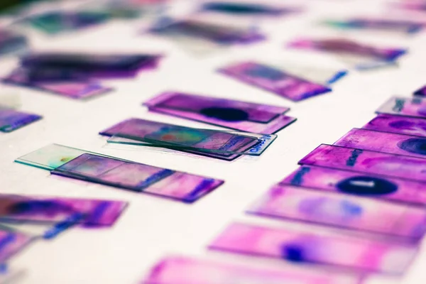
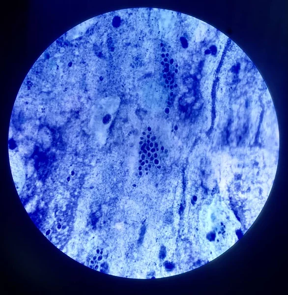
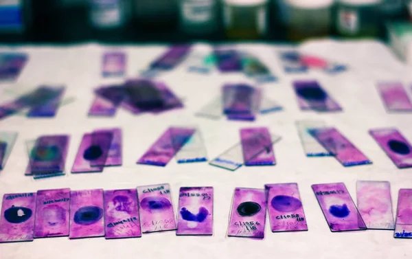
Stained Glass Slides Of Peripheral Blood Smear With Violet Leishman Giemsa Stain In Hematology Pathology Laboratory
Image, 6.66MB, 5184 × 3456 jpg
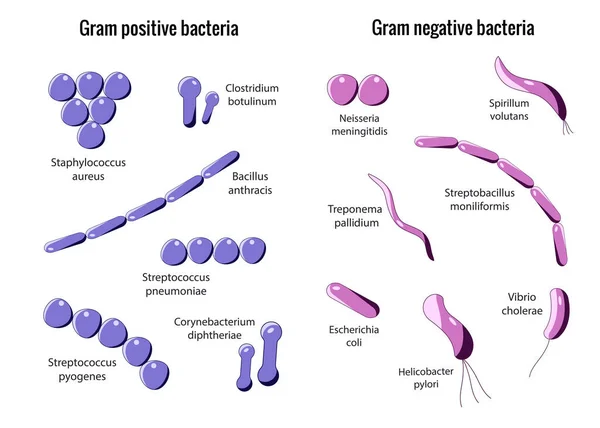
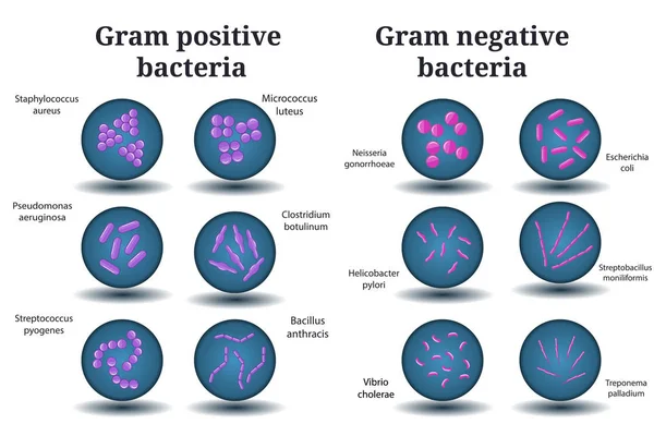
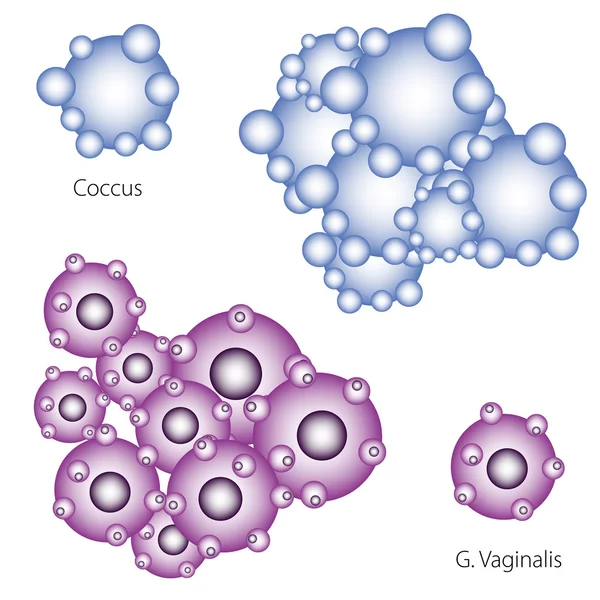
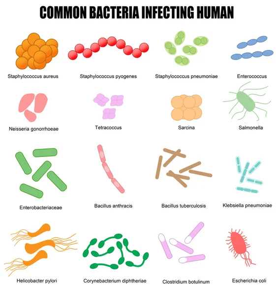
Gram Positive And Gram Negative Bacteria. Coccus, Bacillus, Curved Bacteria In Petri Dish.
Image, 2.22MB, 5001 × 3334 jpg
Gram Staining, Also Called Gram's Method, Is A Method Of Differentiating Bacterial Species Into Two Large Groups (Gram-positive And Gram-negative).
Image, 15.15MB, 4998 × 3594 jpg


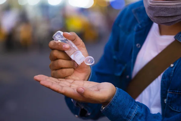
Asian Man Using Wash Hand Sanitizer Gel Dispenser, Against Novel Coronavirus (2019-nCoV) Or Wuhan Coronavirus At Public Train Station Or Supermarket. Antiseptic, Hygiene And Healthcare Concept
Image, 17.09MB, 6000 × 4000 jpg
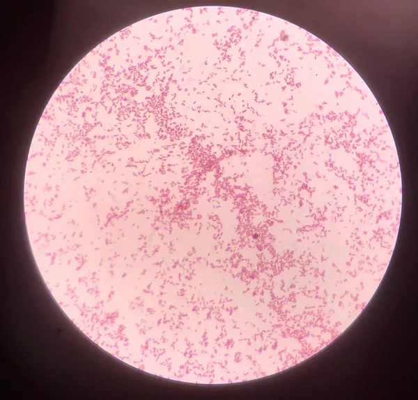

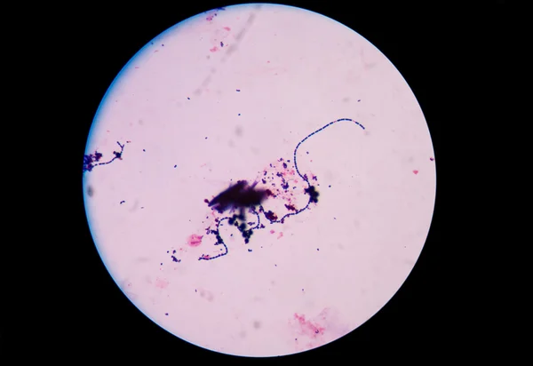
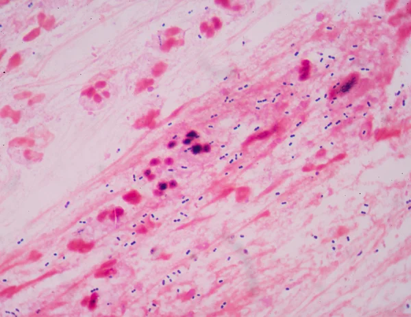
Gram Positive Cocci Bacteria In Gram Staining, Gram Positive Diplococci, Microbiology Lab
Image, 2.34MB, 2176 × 3264 jpg

Gram Positive Cocci Bacteria In Gram Staining, Gram Positive Diplococci, Microbiology Lab
Image, 2.25MB, 2176 × 3264 jpg

Gram Positive Cocci Bacteria In Gram Staining, Gram Positive Diplococci, Microbiology Lab
Image, 2.43MB, 2176 × 3264 jpg


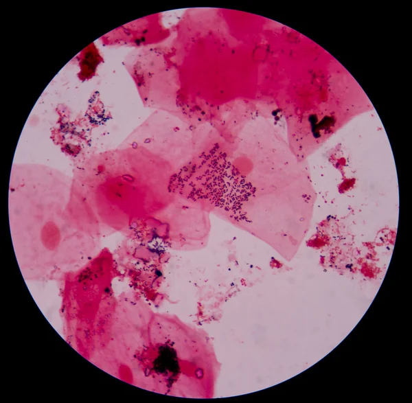


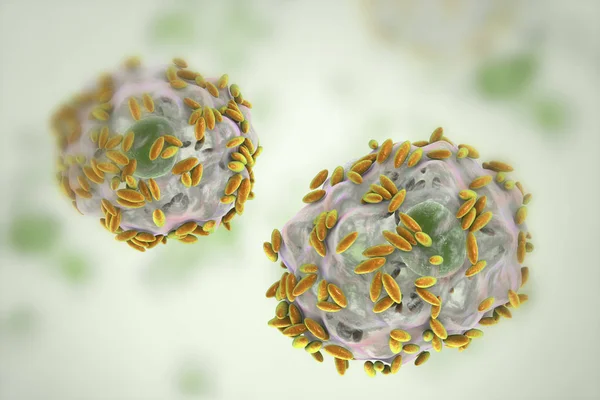
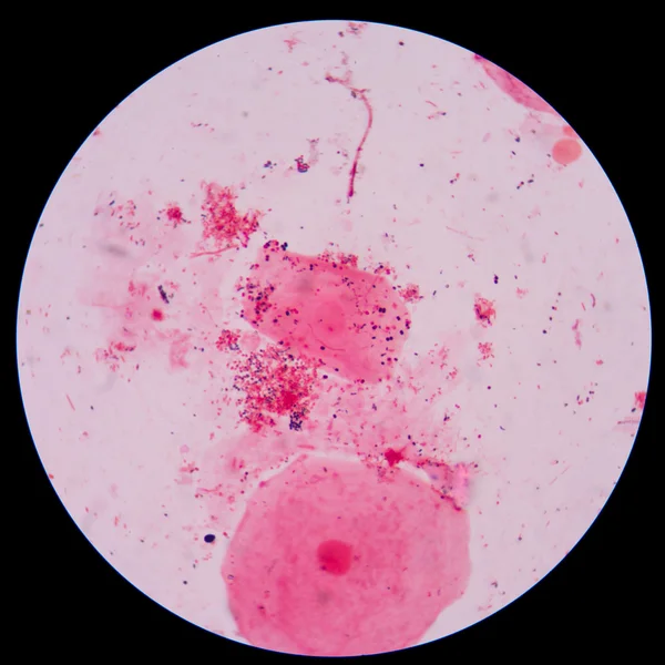
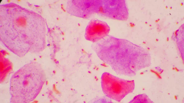
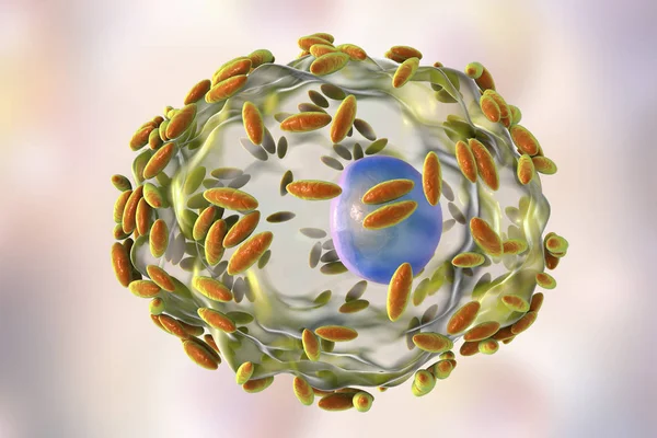
Microscopic Diagnosis Of Bacterial Vaginosis. Epithelial Cell, So-called Clue Cell Is Covered With Bacteria Gardnerella Vaginalis
Image, 3.01MB, 6000 × 4000 jpg
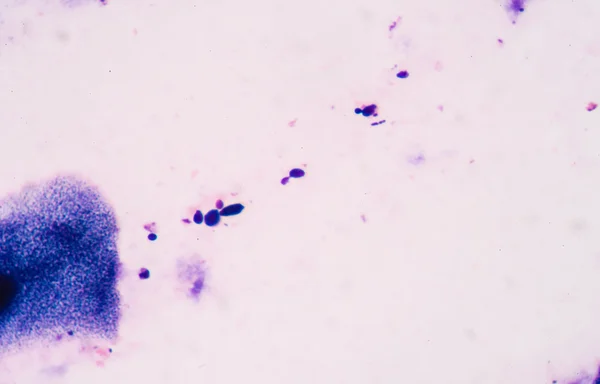
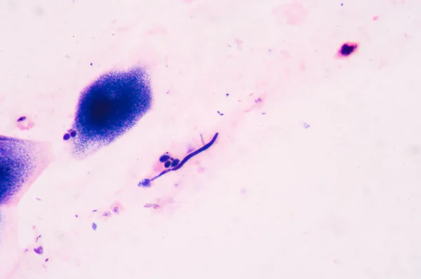
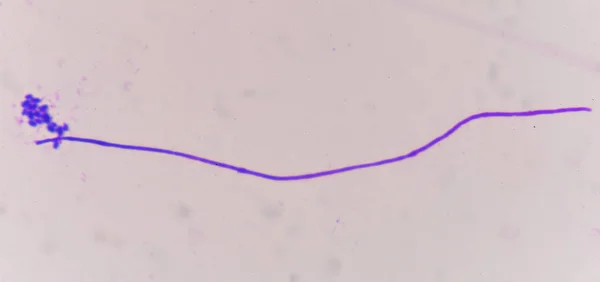
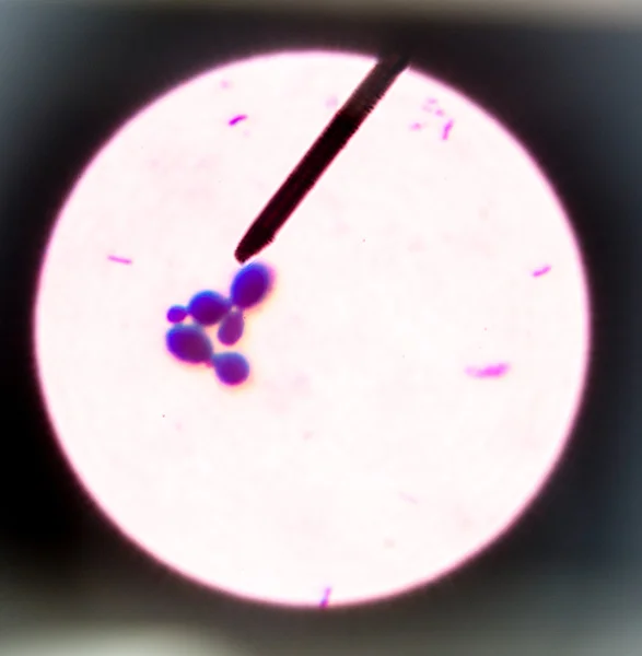
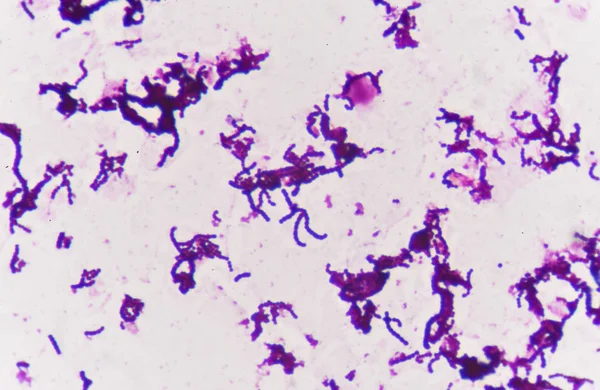
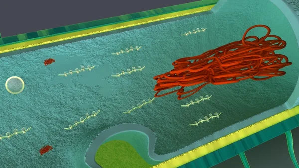
Bacterium Branching Budding Yeast Cells With Pseudohyphae In Urine Gram St
Image, 2.52MB, 2898 × 2897 jpg

Microscopic Diagnosis Of Bacterial Vaginosis. Epithelial Cell, So-called Clue Cell Is Covered With Bacteria Gardnerella Vaginalis
Image, 2.53MB, 6000 × 4000 jpg
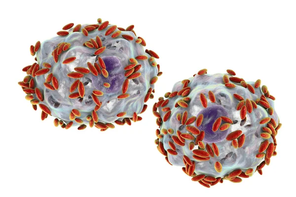
Microscopic Diagnosis Of Bacterial Vaginosis. Epithelial Cell, So-called Clue Cell Is Covered With Bacteria Gardnerella Vaginalis
Image, 3.12MB, 6000 × 4000 jpg
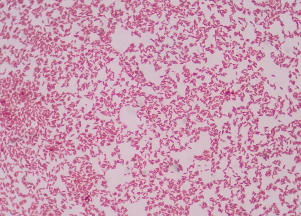
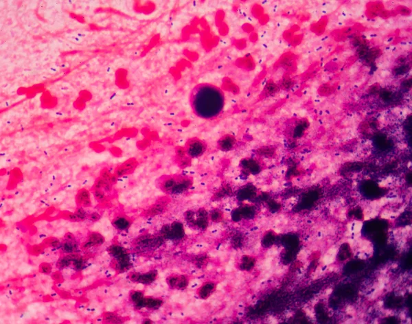
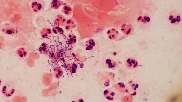
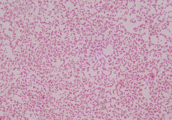
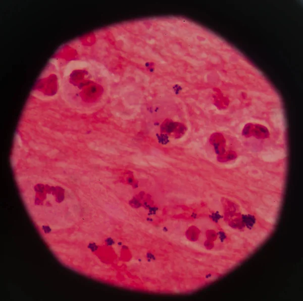
Red Cells Bacteria Cell Gram Neagative Bacilli With Capsule Pathogen.Sample Sputum In Gram Stain Method.
Image, 4.71MB, 4592 × 2584 jpg

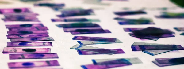
Stained Glass Slides Of Peripheral Blood Smear With Violet Leishman Giemsa Stain In Hematology Pathology Laboratory
Image, 3.83MB, 5183 × 1956 jpg

Stained Glass Slides Of Peripheral Blood Smear With Violet Leishman Giemsa Stain In Hematology Pathology Laboratory
Image, 5.63MB, 5184 × 2991 jpg

Diagram Showing Gram Staining Microbiology Lab Technique Steps - Microbiology Laboratory Using Crystal Violet And Safranin
Vector, 8.54MB, 6001 × 4000 eps

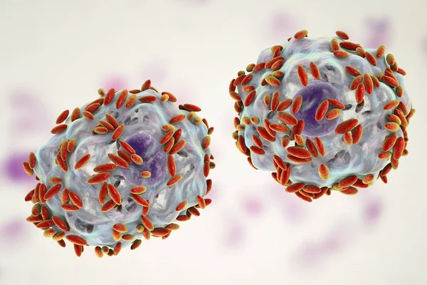
Microscopic Diagnosis Of Bacterial Vaginosis. Epithelial Cell, So-called Clue Cell Is Covered With Bacteria Gardnerella Vaginalis
Image, 3.19MB, 6000 × 4000 jpg
Stained Glass Slides Of Peripheral Blood Smear With Violet Leishman Giemsa Stain In Hematology Pathology Laboratory
Image, 6.3MB, 5183 × 3266 jpg

Microscopic Diagnosis Of Bacterial Vaginosis. Epithelial Cell, So-called Clue Cell Is Covered With Bacteria Gardnerella Vaginalis
Image, 2.63MB, 6000 × 4000 jpg

This Differential Staining Procedure Separates Most Bacteria Into Two Groups On The Basis Of Cell Wall Composition, Bacteria Gram Posotive And Gram Negative
Vector, 11.84MB, 2859 × 6420 eps
Page 1 >> Next