
Stock image Hard Exudates

Non-proliferative Diabetic Retinopathy, 3D Illustration Showing Hard Exudates, Microaneurysms, Dot Haemorrhages, Flame-shaped And Splinter Retinal Haemorrhages, Ophthalmoscope View
Image, 13.11MB, 5352 × 5352 jpg

Non-proliferative Diabetic Retinopathy, 3D Illustration Showing Normal Eye Retina And Retina With Hard Exudates (irregularly Shaped Yellow Spots)
Image, 25.27MB, 11738 × 6603 jpg

Non-proliferative Diabetic Retinopathy, 3D Illustration Showing Normal Eye Retina And Retina With Hard Exudates, And Cotton Wool Spots
Image, 25.26MB, 11738 × 6603 jpg

Non-proliferative Diabetic Retinopathy, Illustration Showing Normal Eye Retina And Retina With Hard Exudates, Microaneurysms, Dot Haemorrhages, Flame-shaped And Splinter Retinal Haemorrhages
Image, 7.35MB, 11738 × 6603 jpg

Non-proliferative Diabetic Retinopathy, Illustration Showing Normal Eye Retina And Retina With Hard Exudates, Microaneurysms, Dot Haemorrhages, Flame-shaped And Splinter Retinal Haemorrhages
Image, 6.89MB, 11738 × 6603 jpg
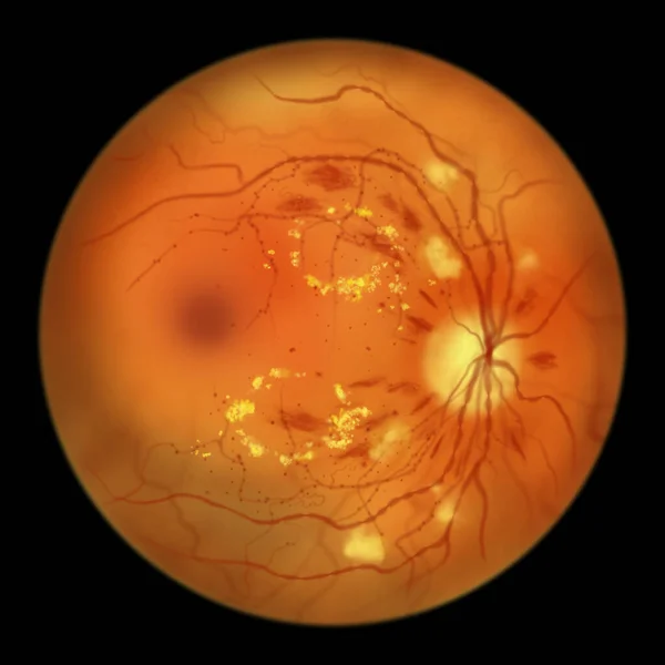
Diabetic Retinopathy Non-proliferative, Illustration Showing Hard Exudates, Cotton Wool Spots, Microaneurysms, Dot Haemorrhages, Flame-shaped And Splinter Retinal Haemorrhages, IRMAs, Venous Beading
Image, 3.07MB, 5000 × 5000 jpg
Proliferative Diabetic Retinopathy, Illustration Showing Neovascularization In The Disk And Other Sites, Macula Edema And Hard Exudates. Fundoscopic Examination Of The Eye Retina In Diabetes Mellitus
Image, 3.05MB, 5000 × 5000 jpg

Diabetic Retinopathy, 3D Illustration Showing Macula Edema, Optic Disk Edema And Hard Exudates, Abnormal Finding On Fundoscopic Examination Of The Eye Retina In Diabetes Mellitus
Image, 26.67MB, 11738 × 6603 jpg

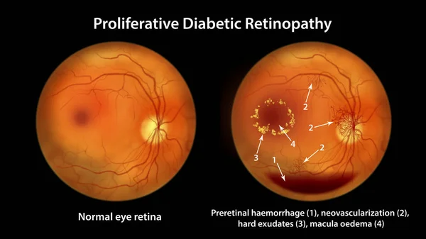
Proliferative Diabetic Retinopathy, 3D Illustration Showing Preretinal Haemorrhage As Horizontal Blood Level, Neovascularization In The Disk And Other Sites, Macula Edema And Hard Exudates
Image, 22.98MB, 11738 × 6603 jpg
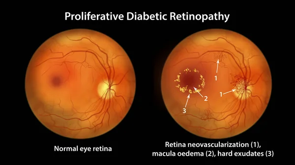
Proliferative Diabetic Retinopathy, Illustration Showing Neovascularization In The Disk And Other Sites, Macula Edema And Hard Exudates. Fundoscopic Examination Of The Eye Retina In Diabetes Mellitus
Image, 6.95MB, 11738 × 6603 jpg
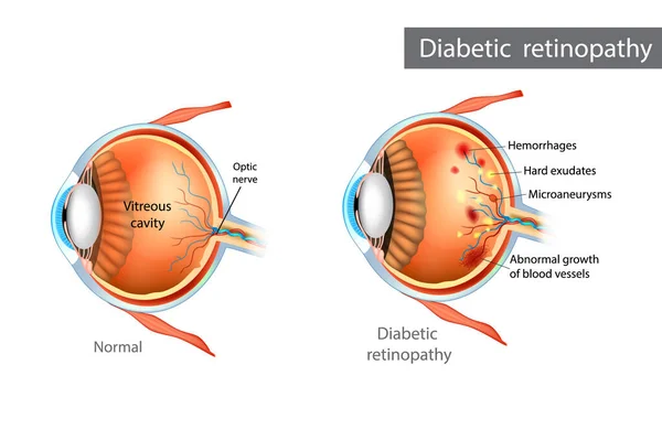
Diabetic Retinopathy. Difference Between Normal Retina And Diabetic Retinopathy
Vector, 2.35MB, 5100 × 3396 eps
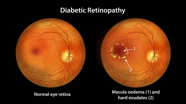
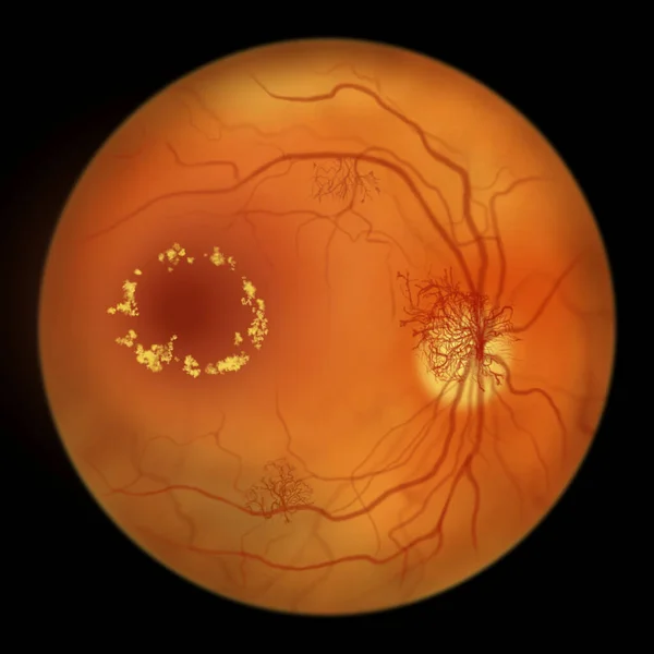
Diabetic Retinopathy, Illustration Showing Macula Edema And Hard Exudates, Abnormal Finding On Fundoscopic Examination Of The Eye Retina In Diabetes Mellitus
Image, 6.56MB, 11738 × 6603 jpg

Diabetic Retinopathy Non-proliferative, Illustration Showing Hard Exudates, Cotton Wool Spots, Microaneurysms, Dot Haemorrhages, Flame-shaped And Splinter Retinal Haemorrhages, IRMAs, Venous Beading
Image, 7.22MB, 11738 × 6603 jpg
Proliferative Diabetic Retinopathy, Illustration Showing Preretinal Haemorrhage As Horizontal Blood Level, Neovascularization In The Disk And Other Sites, Macula Edema And Hard Exudates
Image, 7.07MB, 11738 × 6603 jpg
Page 1 >> Next