Home
Images
Vectors
Videos
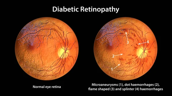
Stock image Non-proliferative diabetic retinopathy, 3D illustration showing normal eye retina and retina with hard exudates (irregularly shaped yellow spots)
Published:
May.10, 2022 06:35:27
Author:
katerynakon
Views:
26
Downloads:
4
File type:
image
/
jpg
File size:
25.27 MB
Orginal size:
11738
x
6603
px
Available sizes:
S
(500 x 281)
M
(1000 x 563)
L
(2000 x 1125)
XL
(11738 x 6603)
EL0
(11738 x 6603)
Level:
silver
Download Image
Image topics:
Illustration
Form
Medicine
Medical
3d
Eye
Clinical
Exam
Spot
Disease
Damaged
Surface
Hard
Diagnosis
Diagnostic
Ophthalmic
Disorder
Ocular
Ophthalmology
Eyeball
Diabetic
Capillary
Leakage
Superficial
Diabetes
Retina
Ophthalmoscopy
Retinal
Capillaries
Endocrinology
Ophthalmoscope
Fundus
Macula
Retinopathy
Exudate
Blood Vessels
Blood Vessel
Diabetic Retinopathy
Diabetes Mellitus
Nonproliferative
Optic Disc
Eye Disease
Retinoscopy
Fundoscopy
Fundus Camera
Funduscopic
Hard Exudates
Similar stock images
Non-proliferative Diabetic Retinopathy, Ophthalmoscope View, 3D Illustration Showing Normal Eye Retina And Retina With Flame-shaped And Splinter Haemorrhages
11738 × 6603
Non-proliferative Diabetic Retinopathy, 3D Illustration Showing Normal Eye Retina And Retina With IRMAs (intraretinal Microvascular Abnormalities) As Small Vessels With Abnormal Branching
11738 × 6603
Non-proliferative Diabetic Retinopathy, Ophthalmoscope View, 3D Illustration Showing Normal Eye Retina And Retina With Multiple Microaneurysms
11738 × 6603
Non-proliferative Diabetic Retinopathy, Ophthalmoscope View, 3D Illustration Showing Normal Eye Retina And Retina With Multiple Dot Haemorrhages (red Dots)
11738 × 6603
Non-proliferative Diabetic Retinopathy, 3D Illustration Showing Normal Eye Retina And Retina With Microaneurysms, Dot Haemorrhages, Flame-shaped And Splinter Retinal Haemorrhages, Ophthalmoscope View
11738 × 6603
Non-proliferative Diabetic Retinopathy, 3D Illustration Showing Normal Eye Retina And Retina With Venous Beading
11738 × 6603
Diabetic Retinopathy, 3D Illustration Shows Preretinal Haemorrhage As Horizontal Blood Level (boat-shaped Haemorrhage), Fundoscopic Examination Of The Eye Retina In Diabetes Mellitus
11738 × 6603
Diabetic Retinopathy Non-proliferative, 3D Illustration Shows Hard Exudates, Cotton Wool Spots, Microaneurysms, Dot Haemorrhages, Flame-shaped And Splinter Retinal Haemorrhages, IRMAs, Venous Beading
11738 × 6603