
Stock image Hematoxylin


Light Microscopic Of Human Ovary Showing Primary And Secondary Follicles. Human Physiology Education.
Image, 26.67MB, 6000 × 4000 jpg


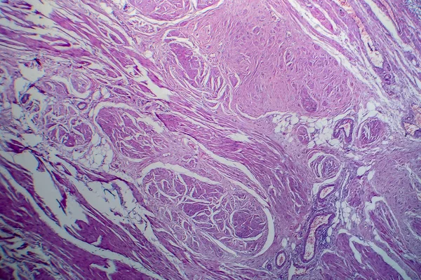
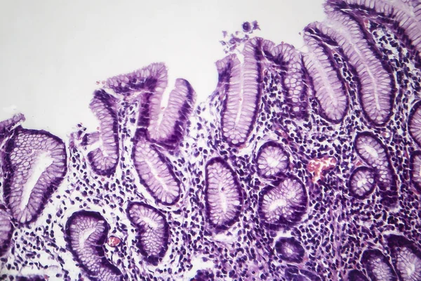
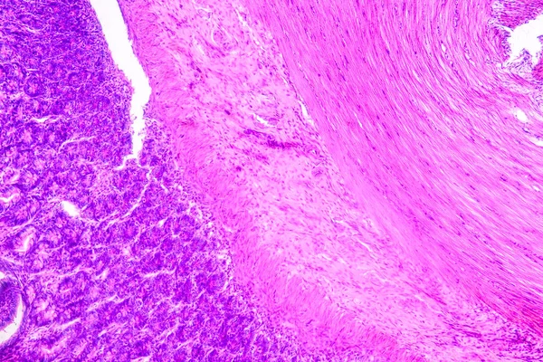
Uterus Adenofibroma, Light Micrograph, Photo Under Microscope. A Rare Benign Tumor Of The Uterus Composed Of Glandular And Fibrous Tissues
Image, 9.96MB, 4602 × 3068 jpg

Chronic Pyelonephritis, Light Micrograph, Photo Under Microscope. High Magnification
Image, 10.19MB, 4832 × 3221 jpg

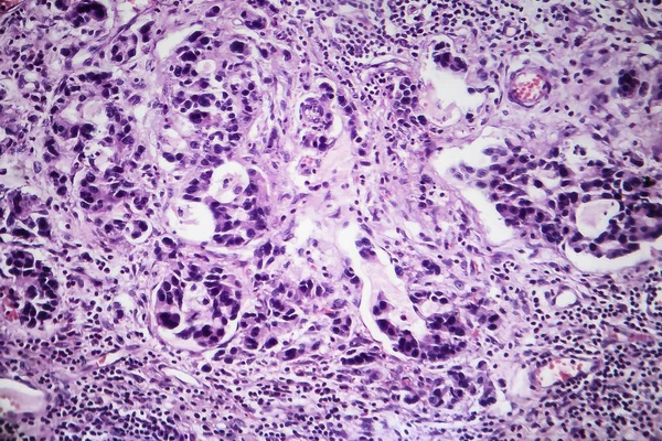
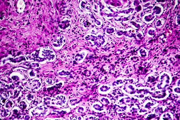
Histopathology Of Diffuse Sclerosing Glomerulonephritis, Light Micrograph, Photo Under Microscope
Image, 8.72MB, 4342 × 2895 jpg
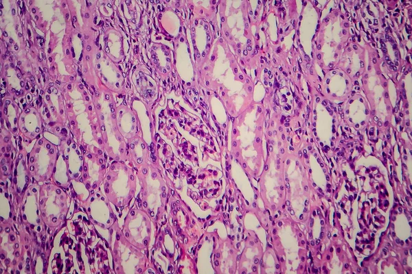
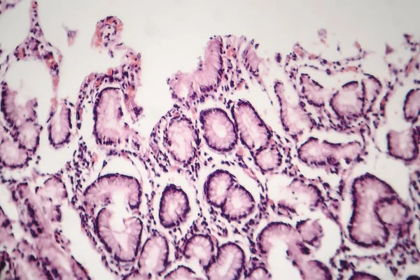
Tubular Atrophy, Light Micrograph, Photo Under Microscope. High Magnification
Image, 7.83MB, 3628 × 2419 jpg
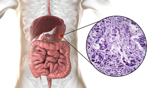
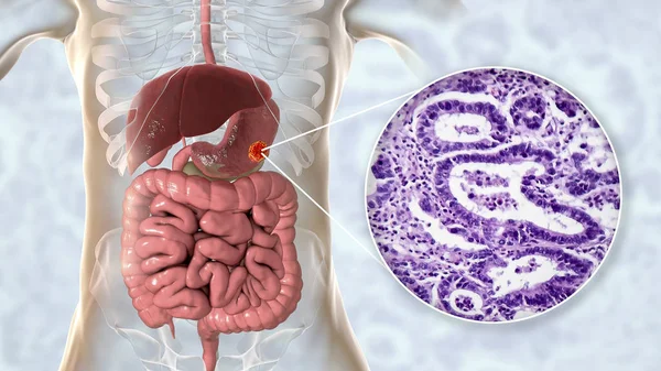
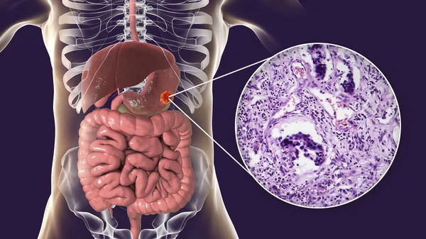
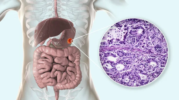
Stomach Adenocarcinoma, Gastric Cancer, Illustration And Light Micrograph
Image, 5.8MB, 5333 × 3000 jpg
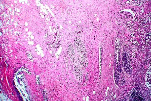
Histopathology Of Interstitial Nephritis, Light Micrograph, Photo Under Microscope
Image, 10.65MB, 3956 × 2637 jpg
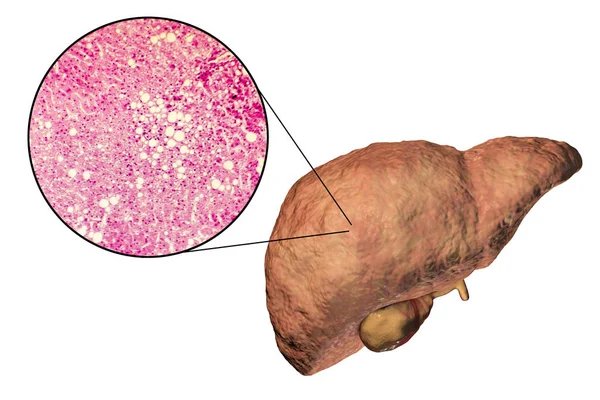
Liver With Cirrhosis Inside Human Body. 3D Illustration And Light Micrograph Of Biliary Cirrhosis
Image, 10.35MB, 5669 × 3189 jpg
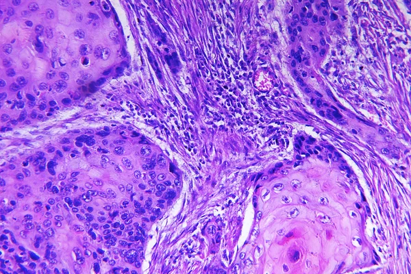
Bladder Transitional Cell Carcinoma, Light Micrograph, Photo Under Microscope
Image, 10.24MB, 4483 × 2989 jpg
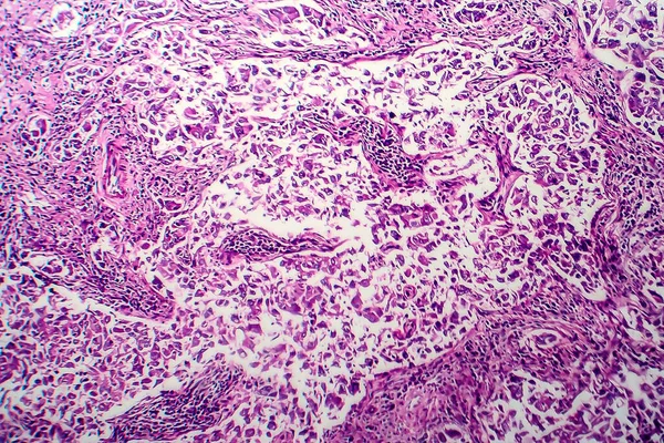
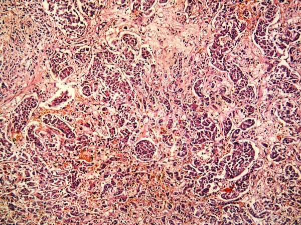
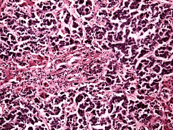
Wilms Tumor, Or Nephroblastoma, Light Micrograph, Photo Under Microscope
Image, 6.57MB, 3582 × 2388 jpg

Stomach Adenocarcinoma, Gastric Cancer, Illustration And Light Micrograph
Image, 6.2MB, 5333 × 3000 jpg
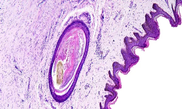
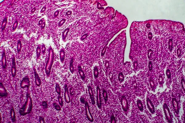
Histology Of Human Tissue, Show Skin With Hair Follicles As Seen Under The Microscope, 10x Zoom
Image, 3.66MB, 2891 × 1739 jpg

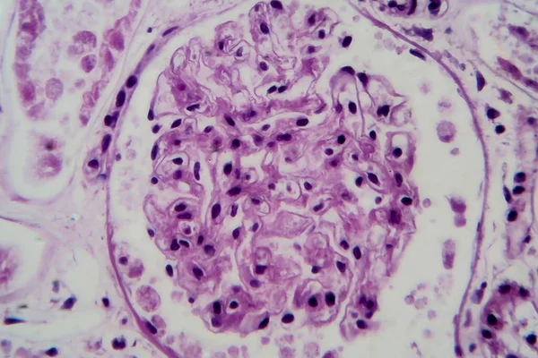
Acute Glomerulonephritis, Light Micrograph, Photo Under Microscope. High Magnification
Image, 5.89MB, 4625 × 3083 jpg

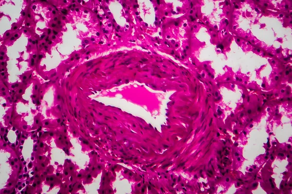
Photomicrograph Of Lung Tissue Depicting Silicosis Pathology Under A Microscope, Revealing Silica Particle Accumulation In Alveoli And Fibrosis.
Image, 26.14MB, 7124 × 4749 jpg
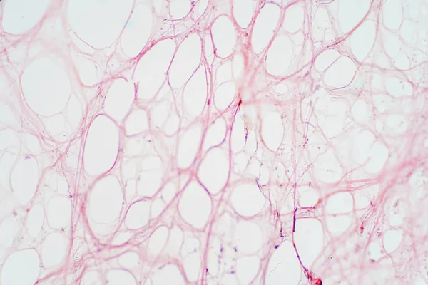
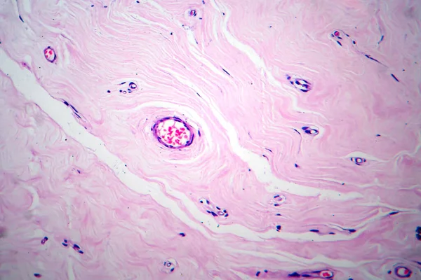
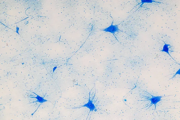
Areolar Connective Tissue Under The Microscope View. Histological For Human Physiology.
Image, 9.51MB, 6000 × 4000 jpg
Stomach Adenocarcinoma, Gastric Cancer, Illustration And Light Micrograph
Image, 6.14MB, 5333 × 3000 jpg
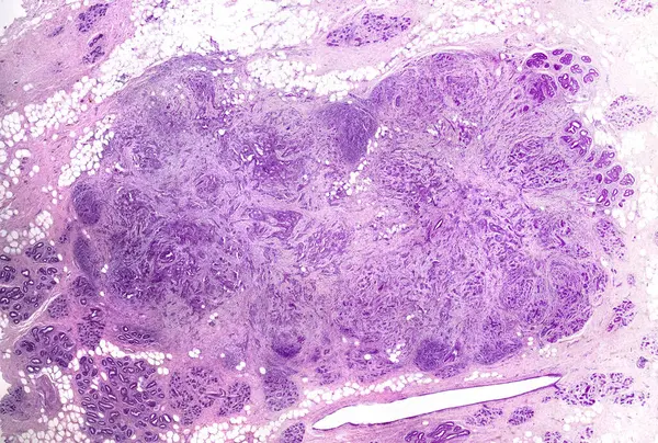
Breast Ductal Carcinoma. Very Low Magnification Light Micrograph Showing Irregular Cords And Nests Of Malignant Ductal Carcinoma Cells Invading Breast Stroma.
Image, 17.99MB, 4554 × 3072 jpg

This Is A Pathological Photo Of Human Left Ventricular Hypertrophy, Showing An Increase In Myocardial Diameter And Interstitial Distance.Magnify 40x.
Image, 42.86MB, 8500 × 8500 jpg
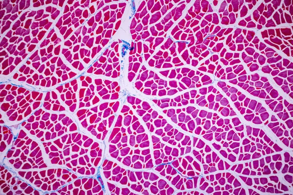
Characteristics Of Anatomy And Histological Sample Striated (Skeletal) Muscle Of Mammal Tissue Under The Microscope.
Image, 29.12MB, 8192 × 5464 jpg


Stomach Adenocarcinoma, Gastric Cancer, Illustration And Light Micrograph
Image, 6.66MB, 5333 × 3000 jpg


Stomach Adenocarcinoma, Gastric Cancer, Illustration And Light Micrograph
Image, 6.46MB, 5333 × 3000 jpg

Light Microscopic Of Human Ovary Showing Primary And Secondary Follicles. Human Physiology Education.
Image, 27.23MB, 6000 × 4000 jpg
Education Anatomy And Histological Sample Of Human Under The Microscope.
Image, 21.93MB, 6720 × 4480 jpg
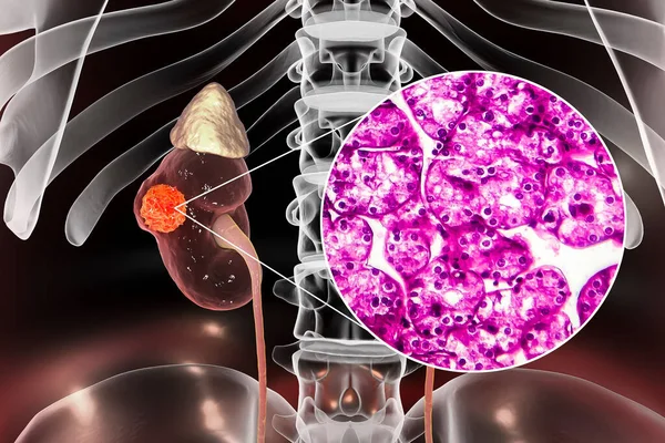
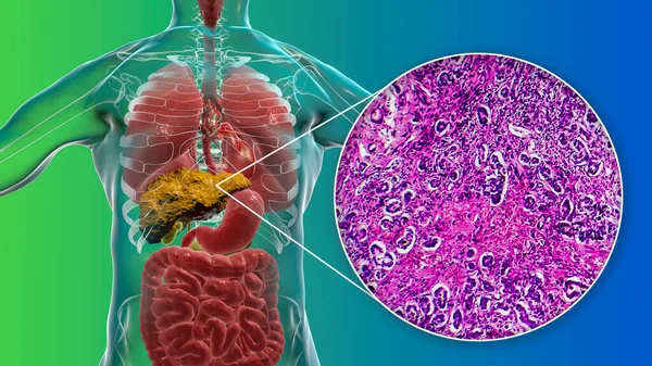
Kidney Cancer, Renal Cell Carcinoma, 3D Illustration And Light Micrograph
Image, 7.28MB, 4500 × 3000 jpg
Page 1 >> Next