
Stock image Hematoxylin page 2

Photomicrograph Of Lung Tissue Depicting Silicosis Pathology Under A Microscope, Revealing Silica Particle Accumulation In Alveoli And Fibrosis.
Image, 26.14MB, 7124 × 4749 jpg
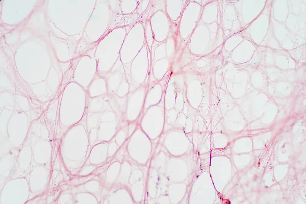
Areolar Connective Tissue Under The Microscope View. Histological For Human Physiology.
Image, 9.51MB, 6000 × 4000 jpg
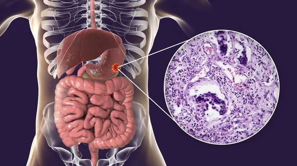
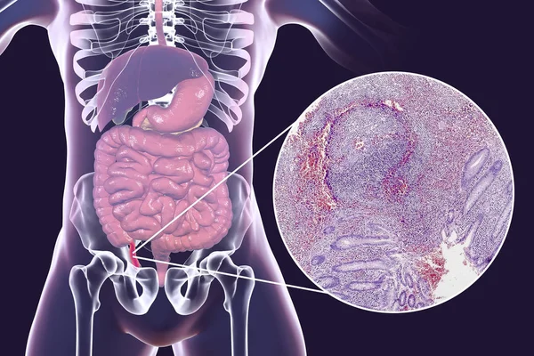
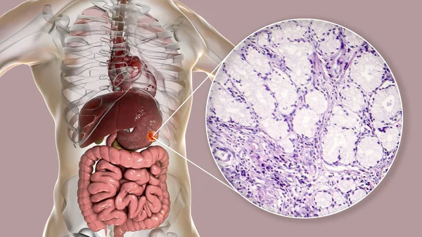
Stomach Adenocarcinoma, Gastric Cancer, Illustration And Light Micrograph
Image, 6.14MB, 5333 × 3000 jpg
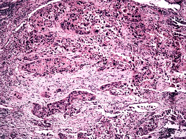
Breast Ductal Carcinoma. Very Low Magnification Light Micrograph Showing Irregular Cords And Nests Of Malignant Ductal Carcinoma Cells Invading Breast Stroma.
Image, 17.99MB, 4554 × 3072 jpg

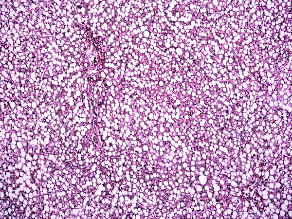
This Is A Pathological Photo Of Human Left Ventricular Hypertrophy, Showing An Increase In Myocardial Diameter And Interstitial Distance.Magnify 40x.
Image, 42.86MB, 8500 × 8500 jpg
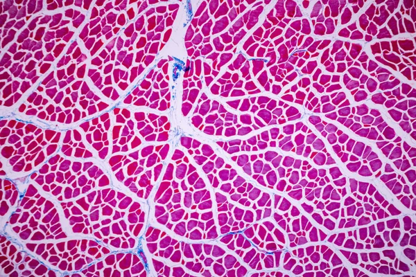
Characteristics Of Anatomy And Histological Sample Striated (Skeletal) Muscle Of Mammal Tissue Under The Microscope.
Image, 29.12MB, 8192 × 5464 jpg

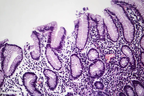

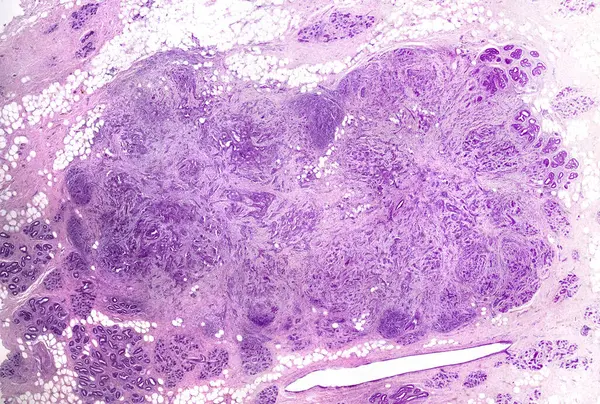
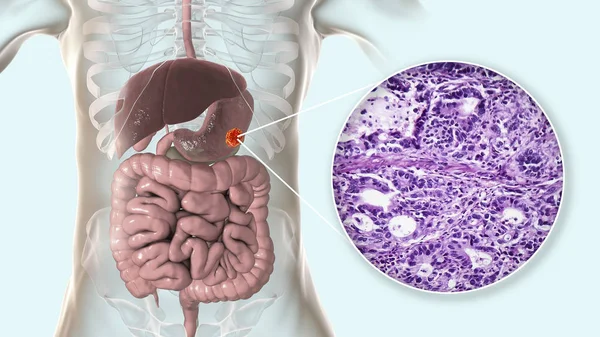
Stomach Adenocarcinoma, Gastric Cancer, Illustration And Light Micrograph
Image, 6.66MB, 5333 × 3000 jpg


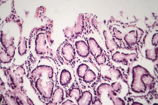
Stomach Adenocarcinoma, Gastric Cancer, Illustration And Light Micrograph
Image, 6.46MB, 5333 × 3000 jpg

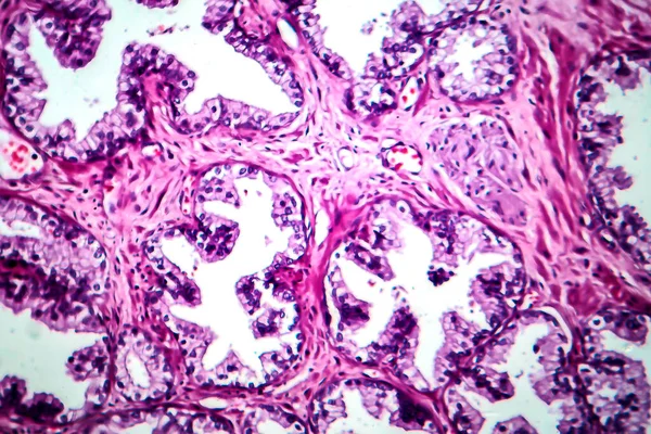
Light Microscopic Of Human Ovary Showing Primary And Secondary Follicles. Human Physiology Education.
Image, 27.23MB, 6000 × 4000 jpg
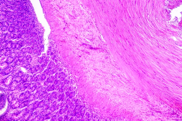
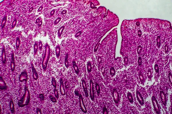
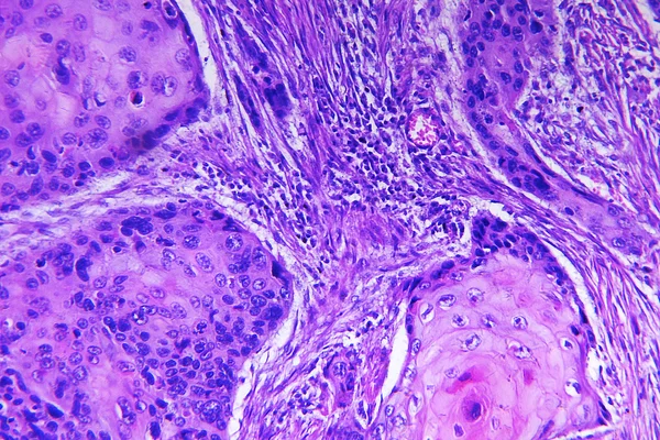
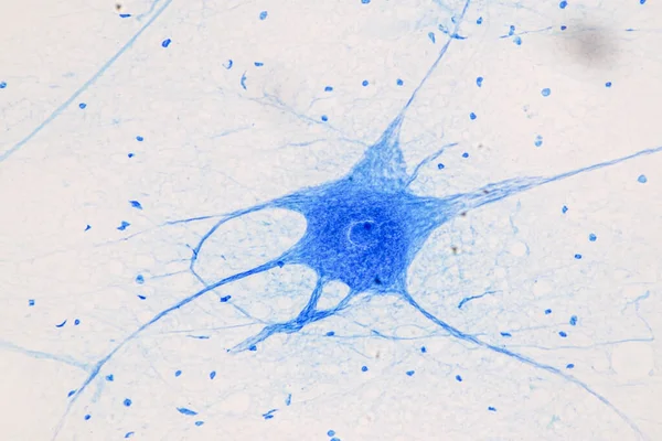
Education Anatomy And Histological Sample Of Human Under The Microscope.
Image, 21.93MB, 6720 × 4480 jpg
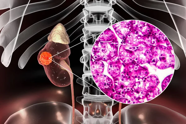
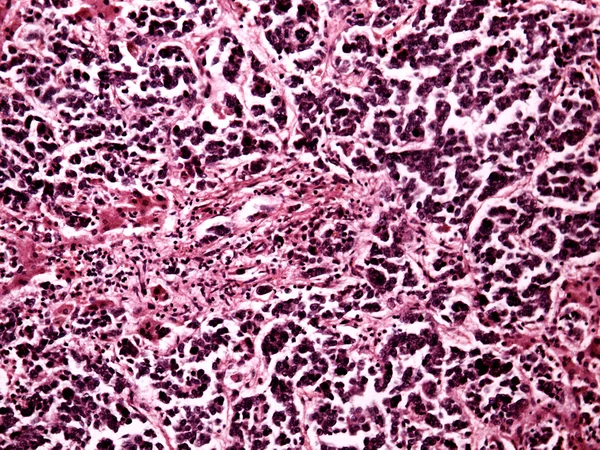
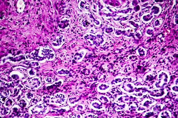
Kidney Cancer, Renal Cell Carcinoma, 3D Illustration And Light Micrograph
Image, 7.28MB, 4500 × 3000 jpg

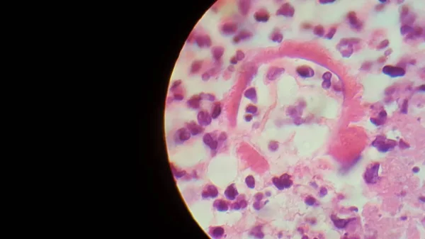
Photomicrograph Of Nasal Polyps, Displaying Abnormal Tissue Growth In The Nasal Passages Often Causing Congestion And Discomfort.
Image, 16.6MB, 6150 × 4100 jpg
Uterine Leiomyoma, Also Known As Fibroids,a Benign Smooth Muscle Tumor Of The Uterus, Light Micrograph, Photo Under Microscope
Image, 15.49MB, 4602 × 3068 jpg
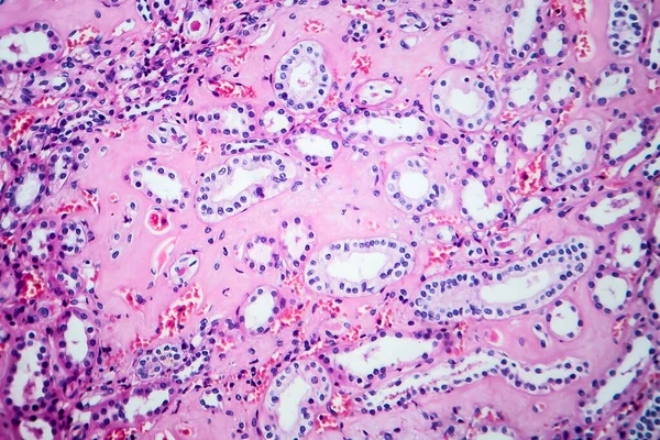
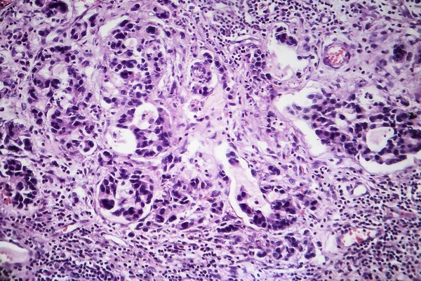
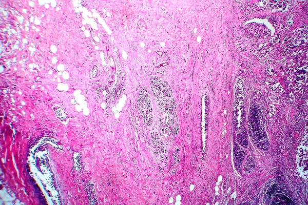
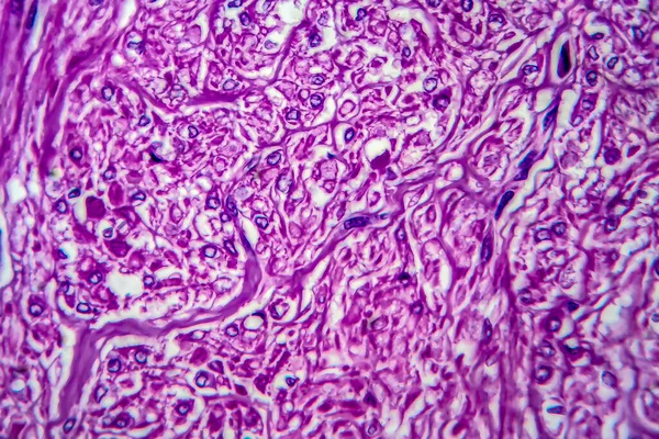
Histopathology Of Hypertensive Renal Disease, Light Micrograph, Photo Under Microscope
Image, 8.81MB, 4657 × 3105 jpg


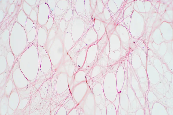
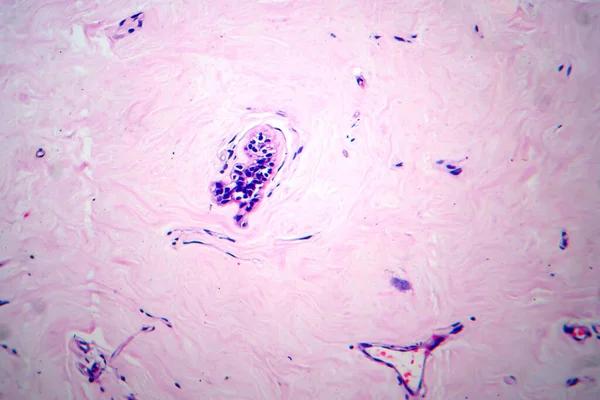
Areolar Connective Tissue Under The Microscope View. Histological For Human Physiology.
Image, 10.06MB, 6000 × 4000 jpg

Very Low Magnification Micrograph Of A Frontal Section Of The Head Of An Embryo Showing, From Top To Bottom, Skin With Developing Hair Bulbs, Nasal Cavities With The Turbinates, Nasal Septum And The Vomeronasal Or Jacobson Organ, Two Teeth Germs, An
Image, 37.47MB, 6739 × 7791 jpg


Uterus Adenofibroma, Light Micrograph, Photo Under Microscope. A Rare Benign Tumor Of The Uterus Composed Of Glandular And Fibrous Tissues
Image, 9.89MB, 4602 × 3068 jpg


Testicular Seminoma, Light Micrograph, Photo Under Microscope. A Most Common Germ Cell Tumor Of The Testis
Image, 15.36MB, 4434 × 2956 jpg
Previous << Page 2 >> Next