
Stock image Histopathology


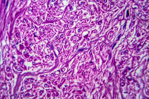
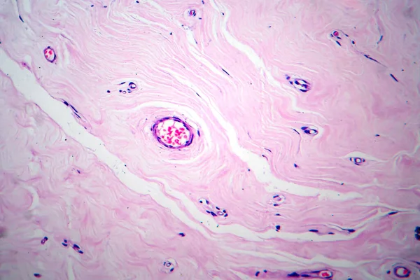
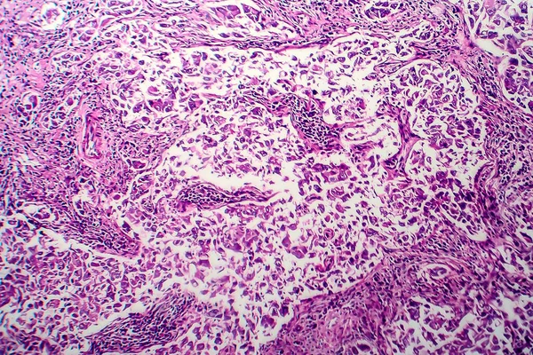
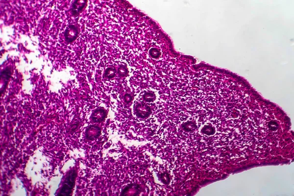
Uterine Leiomyoma, Also Known As Fibroids,a Benign Smooth Muscle Tumor Of The Uterus, Light Micrograph, Photo Under Microscope
Image, 15.49MB, 4602 × 3068 jpg
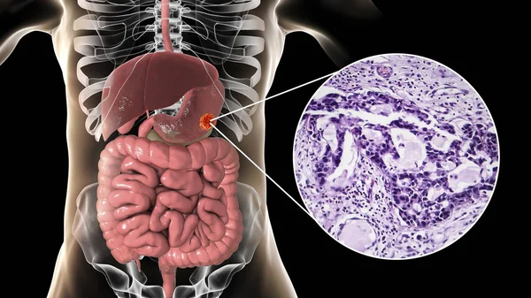
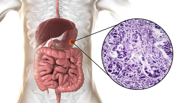
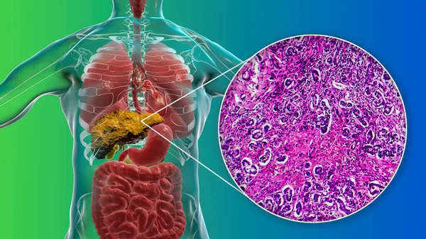
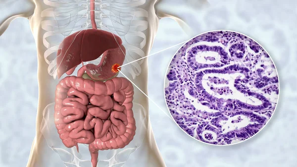
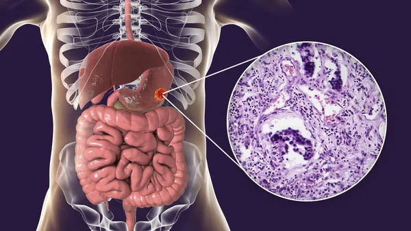
Stomach Adenocarcinoma, Gastric Cancer, Illustration And Light Micrograph
Image, 5.73MB, 5333 × 3000 jpg


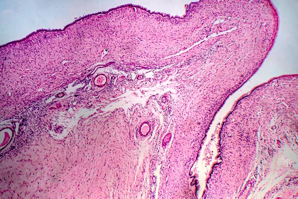
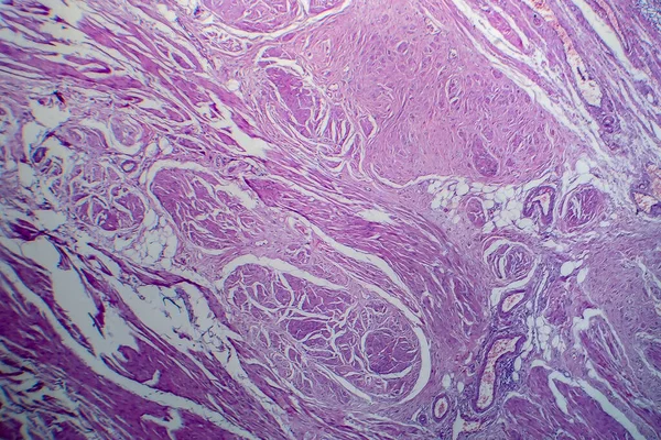
Uterus Adenofibroma, Light Micrograph, Photo Under Microscope. A Rare Benign Tumor Of The Uterus Composed Of Glandular And Fibrous Tissues
Image, 9.96MB, 4602 × 3068 jpg

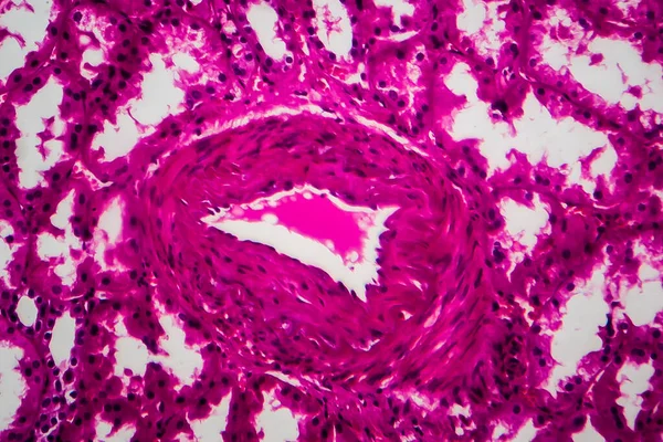
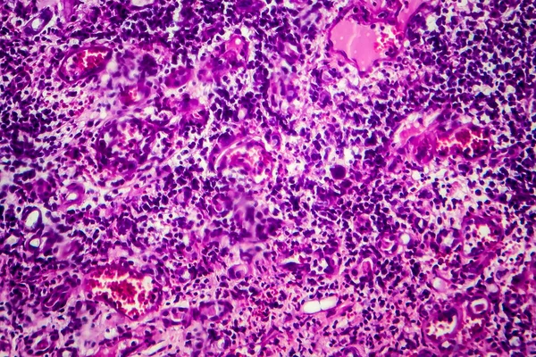
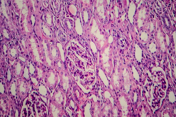
Chronic Pyelonephritis, Light Micrograph, Photo Under Microscope. High Magnification
Image, 10.19MB, 4832 × 3221 jpg

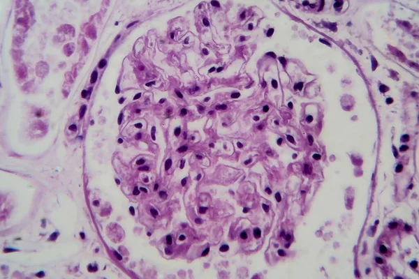
Histopathology Of Diffuse Sclerosing Glomerulonephritis, Light Micrograph, Photo Under Microscope
Image, 8.72MB, 4342 × 2895 jpg
Tubular Atrophy, Light Micrograph, Photo Under Microscope. High Magnification
Image, 7.83MB, 3628 × 2419 jpg
Stomach Adenocarcinoma, Gastric Cancer, Illustration And Light Micrograph
Image, 5.8MB, 5333 × 3000 jpg
Histopathology Of Interstitial Nephritis, Light Micrograph, Photo Under Microscope
Image, 10.65MB, 3956 × 2637 jpg
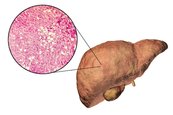
Liver With Cirrhosis Inside Human Body. 3D Illustration And Light Micrograph Of Biliary Cirrhosis
Image, 10.35MB, 5669 × 3189 jpg
Bladder Transitional Cell Carcinoma, Light Micrograph, Photo Under Microscope
Image, 10.24MB, 4483 × 2989 jpg
Wilms Tumor, Or Nephroblastoma, Light Micrograph, Photo Under Microscope
Image, 6.57MB, 3582 × 2388 jpg

Stomach Adenocarcinoma, Gastric Cancer, Illustration And Light Micrograph
Image, 6.2MB, 5333 × 3000 jpg

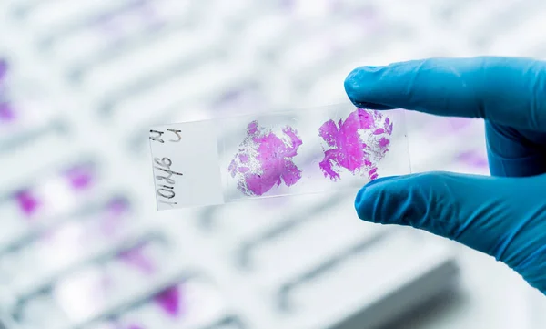
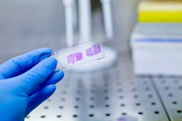
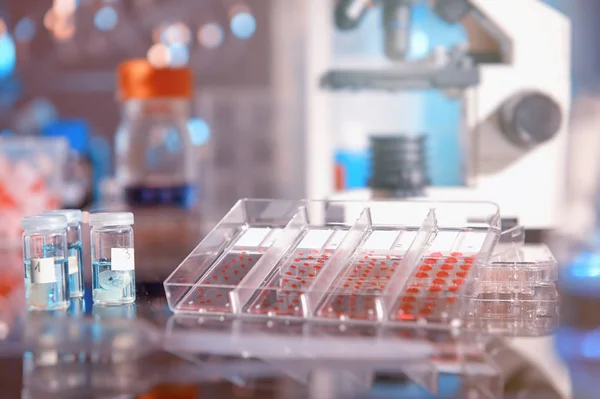
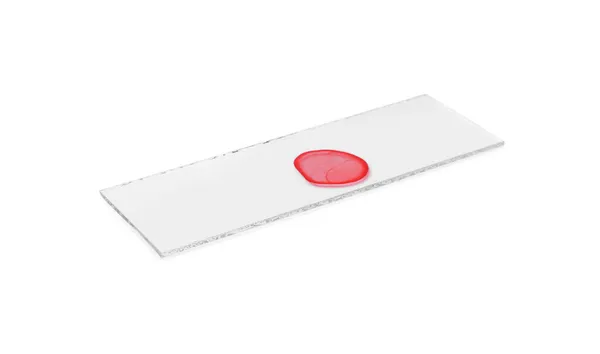
Modern Science Background. Hand In Blue Glove With Sample, Working Scientists Out Of Focus, Text Space
Image, 7.89MB, 5599 × 3200 jpg

Acute Glomerulonephritis, Light Micrograph, Photo Under Microscope. High Magnification
Image, 5.89MB, 4625 × 3083 jpg
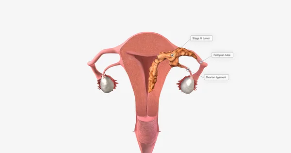
During Stage III Endometrial Cancer, The Tumor Spreads Outside Of The Uterus. 3D Rendering
Image, 1.86MB, 7340 × 3884 jpg
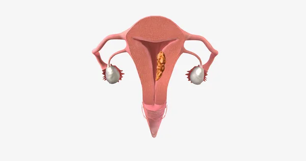
Stage II Endometrial Cancer Is Characterized By Tumor Spread To The Uterine Cervix. 3D Rendering
Image, 1.65MB, 7340 × 3884 jpg

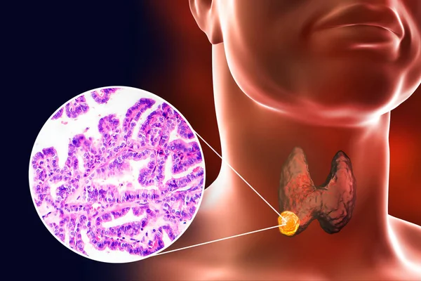
A 3D Scientific Illustration Showcasing A Human Body With Transparent Skin, Revealing A Tumor In His Thyroid Gland, Along With A Micrograph Image Of Papillary Thyroid Carcinoma.
Image, 7.63MB, 6750 × 4500 jpg

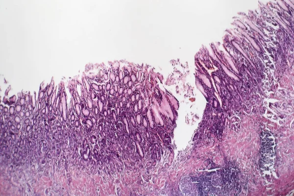
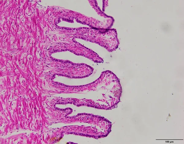
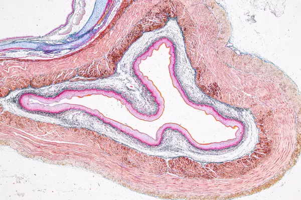
This Is A Histological Photograph Of The Human Small Intestine. Magnify 40x.
Image, 9.15MB, 4202 × 4202 jpg
Stomach Adenocarcinoma, Gastric Cancer, Illustration And Light Micrograph
Image, 6.14MB, 5333 × 3000 jpg

Humanoid Robot Working With Microscope, Conceptual 3D Illustration. Laboratory Automation. Artificial Intelligence For Medicine, Science And Laboratory Industry
Image, 6.13MB, 6400 × 4800 jpg

Pathology And Histology Tissue Of Mouse, Rabbit, Cat And Cow Under Microscope.
Image, 32.75MB, 6000 × 4000 jpg

Pathology And Histology Tissue Of Mouse, Rabbit, Cat And Cow Under Microscope.
Image, 6.85MB, 2667 × 4000 jpg
Pathology And Histology Tissue Of Mouse, Rabbit, Cat And Cow Under Microscope.
Image, 18.37MB, 6000 × 4000 jpg

Pathology And Histology Tissue Of Mouse, Rabbit, Cat And Cow Under Microscope.
Image, 8.03MB, 6000 × 3245 jpg

Oncologist, Pet Veterinarian Examines An MRI Of A Cat For A Tumor And Finds One. Note: This Cat Has Had A Mastectomy For Cancer. Veterinary Medicine.
Image, 2.21MB, 4715 × 3027 jpg

A Man With Lung Cancer, 3D Illustration, Along With A Micrograph Image Of Small Cell Lung Cancer.
Image, 13.35MB, 6000 × 4000 jpg


Page 1 >> Next