photo free
Home Images Vectors Videos

Stock image Immune System Response

stock image White blood cells overview

White Blood Cells Overview

Image, 5.68MB, 9375 × 6250 jpg
stock image Bacteria attacking immune system

Bacteria Attacking Immune System

Image, 4.18MB, 3840 × 2160 jpg
stock image Detailed 3D illustration revealing the intricate inner structure of a monocyte cell, vital in the immune system's defense.

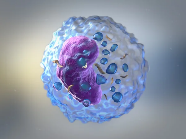
Detailed 3D Illustration Revealing The Intricate Inner Structure Of A Monocyte Cell, Vital In The Immune System's Defense.

Image, 6.11MB, 7200 × 4050 jpg
stock vector HPV infection abstract concept vector illustrations.

HPV Infection Abstract Concept Vector Illustrations.

Vector, 6.8MB, 7292 × 3126 eps
stock vector Activation of B-cell leukocytes

Activation Of B-cell Leukocytes

Vector, 8.23MB, 4799 × 4799 eps
stock vector Cancer and cytotoxic T-cells. T lymphocyte kills cancer cells. T-cell (immune responses), release the perforin and granzymes, and attack cancerous cells. Through the action of perforin, granzymes enter the cytoplasm of the target cell, and lead to ap

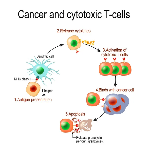
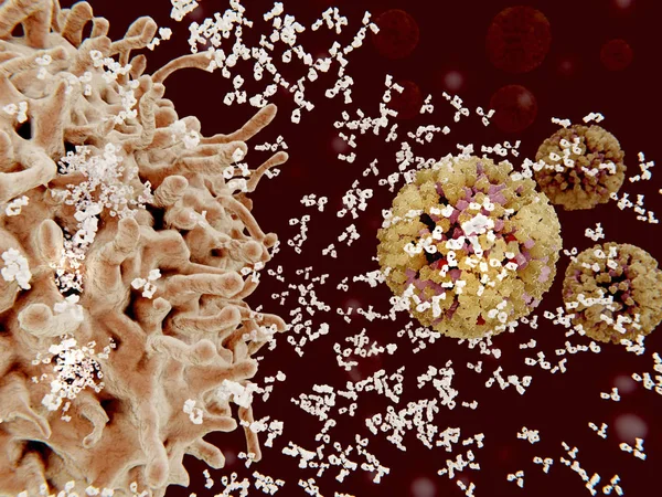
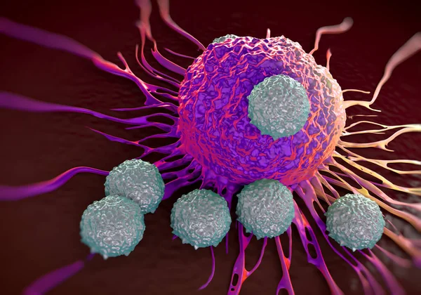
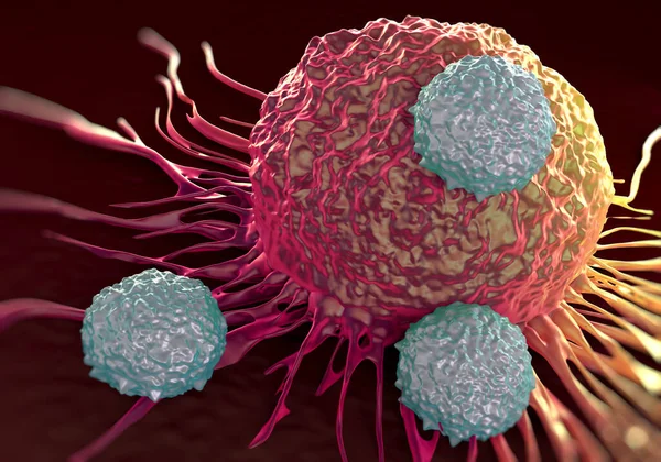
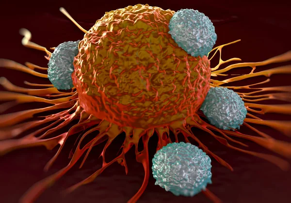
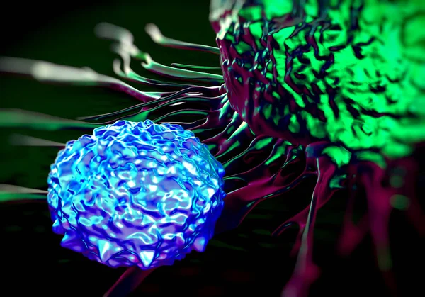
Cancer And Cytotoxic T-cells. T Lymphocyte Kills Cancer Cells. T-cell (immune Responses), Release The Perforin And Granzymes, And Attack Cancerous Cells. Through The Action Of Perforin, Granzymes Enter The Cytoplasm Of The Target Cell, And Lead To Ap

Vector, 1.01MB, 4050 × 4049 eps
stock image Makrophage engulfing an destroying coronaviruses.

Makrophage Engulfing An Destroying Coronaviruses.

Image, 12.3MB, 8000 × 6000 jpg
stock vector T-Cell activation diagram, vector scheme illustration.

T-Cell Activation Diagram, Vector Scheme Illustration.

Vector, 3.21MB, 5734 × 4909 eps
stock image Plasma cells (B-cells) segregate specific antibodies to mark an subsequently destroy viruses (influenza viruses).3D rendering. Illustration

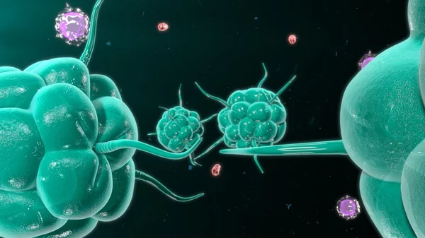
Plasma Cells (B-cells) Segregate Specific Antibodies To Mark An Subsequently Destroy Viruses (influenza Viruses).3D Rendering. Illustration

Image, 9.74MB, 8000 × 6000 jpg
stock vector B-cells and T-cells schematic diagram, vector illustration.

B-cells And T-cells Schematic Diagram, Vector Illustration.

Vector, 3.72MB, 5734 × 5660 eps
stock image Bacteria attacking immune system

Bacteria Attacking Immune System

Image, 4.18MB, 3840 × 2160 jpg
stock vector Blood cells. Set of colored cells. Red blood cells, platelets, leukocytes, lymphocytes, eosinophils, neutrophils, basophils, monocytes. Infographics. Vector illustration on isolated background

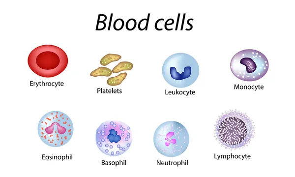
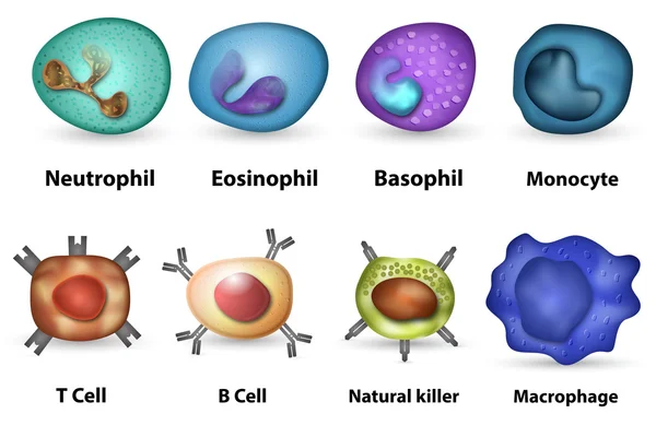
Blood Cells. Set Of Colored Cells. Red Blood Cells, Platelets, Leukocytes, Lymphocytes, Eosinophils, Neutrophils, Basophils, Monocytes. Infographics. Vector Illustration On Isolated Background

Vector, 10.4MB, 6500 × 3900 eps
stock image red blood cells with white blood cells background.

Red Blood Cells With White Blood Cells Background.

Image, 4.87MB, 5157 × 3617 jpg
stock vector Stem cell. The development of red blood cells, leukocytes, macrophages, lymphocytes  and platelets. Infographics.

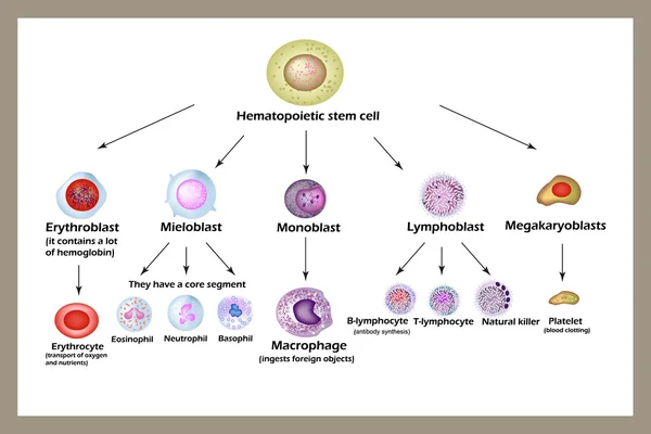
Stem Cell. The Development Of Red Blood Cells, Leukocytes, Macrophages, Lymphocytes And Platelets. Infographics.

Vector, 10MB, 5000 × 3333 eps
stock image Lymphocytes are white blood cells or leucocytes in the human imm

Lymphocytes Are White Blood Cells Or Leucocytes In The Human Imm

Image, 3.43MB, 5330 × 4000 jpg
stock image Lung Allergy Concept

Lung Allergy Concept

Image, 8.75MB, 3955 × 3949 jpg
stock image T-cells attacking cancer cell  illustration of  microscopic photos

T-cells Attacking Cancer Cell Illustration Of Microscopic Photos

Image, 1.39MB, 5000 × 3500 jpg
stock image T-cells attacking cancer cell  illustration of  microscopic photos

T-cells Attacking Cancer Cell Illustration Of Microscopic Photos

Image, 1.32MB, 5000 × 3500 jpg
stock image Bacteria attacking immune system

Bacteria Attacking Immune System

Image, 2.04MB, 3840 × 2160 jpg
stock image T-cells attacking cancer cell  illustration of  microscopic photos

T-cells Attacking Cancer Cell Illustration Of Microscopic Photos

Image, 1.25MB, 5000 × 3500 jpg
stock image neutrophil and lymphocyte

Neutrophil And Lymphocyte

Image, 4.11MB, 3872 × 2592 jpg
stock image Plasma cells (B-cells) segregate antibodies to mark to mark an subsequently destroy viruses (influenza viruses).

Plasma Cells (B-cells) Segregate Antibodies To Mark To Mark An Subsequently Destroy Viruses (influenza Viruses).

Image, 7.85MB, 8000 × 6000 jpg
stock image neutrophil and lymphocyte

Neutrophil And Lymphocyte

Image, 5.06MB, 3872 × 2592 jpg
stock image Eosinophil

Eosinophil

Image, 4.82MB, 3872 × 2592 jpg
stock image A microglia cell. It plays an important role in the pathogenesis of Alzheimer's disease

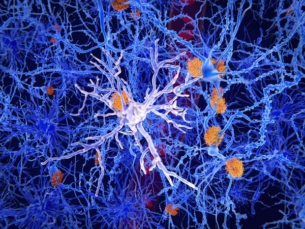
A Microglia Cell. It Plays An Important Role In The Pathogenesis Of Alzheimer's Disease

Image, 13.48MB, 8000 × 6000 jpg
stock image Immune response to coronavirus infection: coronaviruses marked bei antibodies are engulfed and destroyed by a macrophage immune cell

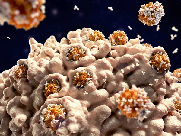
Immune Response To Coronavirus Infection: Coronaviruses Marked Bei Antibodies Are Engulfed And Destroyed By A Macrophage Immune Cell

Image, 8.05MB, 8000 × 6000 jpg
stock image Lymphocyte

Lymphocyte

Image, 6MB, 3872 × 2592 jpg
stock image neutrophil

Neutrophil

Image, 4.27MB, 3872 × 2592 jpg
stock image Neutrophil

Neutrophil

Image, 3.39MB, 3872 × 2592 jpg
stock image Eosinophil

Eosinophil

Image, 4.09MB, 3872 × 2592 jpg
stock image T cells and B cells in immune system

T Cells And B Cells In Immune System

Image, 2.47MB, 3840 × 2160 jpg
stock image Neutrophil

Neutrophil

Image, 4.31MB, 3872 × 2592 jpg
stock image metamyelocyte

Metamyelocyte

Image, 4.98MB, 3872 × 2592 jpg
stock image Lymphoblast

Lymphoblast

Image, 4.54MB, 3872 × 2592 jpg
stock image neutrophil

Neutrophil

Image, 4.58MB, 3872 × 2592 jpg
stock image neutrophil

Neutrophil

Image, 4.4MB, 3872 × 2592 jpg
stock image Neutrophil&Basophil

Neutrophil&Basophil

Image, 5.52MB, 3872 × 2592 jpg
stock image Neutrophil

Neutrophil

Image, 5.27MB, 3872 × 2592 jpg
stock image Neutrophil&Eosinophil

Neutrophil&Eosinophil

Image, 5.17MB, 3872 × 2592 jpg
stock image lymphoblaste

Lymphoblaste

Image, 5.32MB, 3872 × 2592 jpg
stock image COVID-19 vaccine: SARS-CoV-2 spike protein is stabilised for it's use as a vaccine in a tightly closed prefusion conformation through polysorbate (violet) and fatty acid (not visible) molecules. In blue, N-acetylglucosamine. Source: PDB entry 7e7b.

COVID-19 Vaccine: SARS-CoV-2 Spike Protein Is Stabilised For It's Use As A Vaccine In A Tightly Closed Prefusion Conformation Through Polysorbate (violet) And Fatty Acid (not Visible) Molecules. In Blue, N-acetylglucosamine. Source: PDB Entry 7e7b.

Image, 7.43MB, 8000 × 6000 jpg
stock image Basophil

Basophil

Image, 4.93MB, 3872 × 2592 jpg
stock image T-cells attacking cancer cell  illustration of  microscopic photos

T-cells Attacking Cancer Cell Illustration Of Microscopic Photos

Image, 1.33MB, 5000 × 3500 jpg
stock image lymphocyte

Lymphocyte

Image, 4.8MB, 3872 × 2592 jpg
stock image Eosinophil

Eosinophil

Image, 4.53MB, 3872 × 2592 jpg
stock image lymphoblaste

Lymphoblaste

Image, 6.14MB, 3872 × 2592 jpg
stock image The T-cell receptor activates the immune response to antigens in T-lymphocytes. T-cell receptors (dark blue), CD4 molecules (light blue), glycolipids (orange). 3d rendering. Illustration

The T-cell Receptor Activates The Immune Response To Antigens In T-lymphocytes. T-cell Receptors (dark Blue), CD4 Molecules (light Blue), Glycolipids (orange). 3d Rendering. Illustration

Image, 3.11MB, 8000 × 6000 jpg
stock image Eosinophil

Eosinophil

Image, 4.42MB, 3872 × 2592 jpg
stock image Lymphoblast

Lymphoblast

Image, 5.36MB, 3872 × 2592 jpg
stock image Ribosome translating mRNA into a polypeptide chain

Ribosome Translating MRNA Into A Polypeptide Chain

Image, 8.4MB, 8000 × 6000 jpg
Page 1 >> Next